当前位置:
X-MOL 学术
›
J. Morphol.
›
论文详情
Our official English website, www.x-mol.net, welcomes your feedback! (Note: you will need to create a separate account there.)
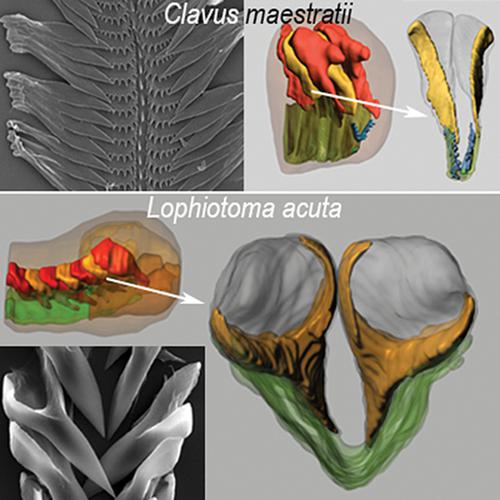
Radula formation in two species of Conoidea (Gastropoda)
Journal of Morphology ( IF 1.5 ) Pub Date : 2020-08-24 , DOI: 10.1002/jmor.21250 Elena Vortsepneva 1 , David G Herbert 2 , Yuri Kantor 3
Journal of Morphology ( IF 1.5 ) Pub Date : 2020-08-24 , DOI: 10.1002/jmor.21250 Elena Vortsepneva 1 , David G Herbert 2 , Yuri Kantor 3
Affiliation
|
The radula is the basic feeding structure in gastropod molluscs and exhibits great morphological diversity that reflects the exceptional anatomical and ecological diversity occurring in these animals. This uniquely molluscan structure is formed in the blind end of the radular sac by specialized cells (membranoblasts and odontoblasts). Secretion type, and the number and shape of the odontoblasts that form each tooth characterize the mode of radula formation. These characteristics vary in different groups of gastropods. Elucidation of this diversity is key to identifying the main patterns of radula formation in Gastropoda. Of particular interest would be a phylogenetically closely related group that is characterized by high variability of the radula. One such group is the large monophyletic superfamily Conoidea, the radula of which is highly variable and may consist of the radular membrane with five teeth per row, or the radular membrane with only two or three teeth per row, or even just two harpoon‐like teeth per row without a radular membrane. We studied the radulae of two species of Conoidea (Clavus maestratii Kilburn, Fedosov & Kantor, 2014 [Drilliidae] and, Lophiotoma acuta (Perry, 1811) [Turridae]) using light and electron microscopy. Based on these data and previous studies, we identify the general patterns of the radula formation for all Conoidea: the dorsolateral position of two groups of odontoblasts, uniform size, and shape of odontoblasts, folding of the radula in the radular sac regardless of the radula configuration. The morphology of the subradular epithelium is most likely adaptive to the radula type.
中文翻译:

两种Conoidea(Gastropoda)中的Radula形成
齿叶是腹足类软体动物的基本摄食结构,表现出巨大的形态多样性,反映了这些动物异常的解剖学和生态学多样性。这种独特的软体动物结构是由专门的细胞(成膜细胞和成牙本质细胞)在放射状囊的盲端形成的。分泌物类型以及形成每颗牙齿的成牙本质细胞的数量和形状表征了齿槽的形成模式。这些特征在不同的腹足动物群中有所不同。阐明这种多样性是确定腹足纲中齿叶形成主要模式的关键。特别令人感兴趣的是一个系统发育密切相关的群体,其特征是齿舌的高度可变性。一个这样的群体是大的单系超科Conoidea,其轮齿变化很大,可能由每行五个齿的轮状膜组成,或者每行只有两个或三个齿的轮状膜,甚至每行只有两个鱼叉状的齿而没有轮状膜。我们使用光学和电子显微镜研究了两种 Conoidea (Clavus maestratii Kilburn, Fedosov & Kantor, 2014 [Drilliidae] 和 Lophiotoma acuta (Perry, 1811) [Turridae]) 的轮叶。基于这些数据和以前的研究,我们确定了所有锥状体的齿状突起形成的一般模式:两组成牙本质细胞的背外侧位置、均匀的大小和成牙本质细胞的形状、齿状突起在齿状突起囊中的折叠与齿状突起无关配置。桡骨下上皮的形态最有可能适应于轮齿类型。
更新日期:2020-08-24
中文翻译:
两种Conoidea(Gastropoda)中的Radula形成
齿叶是腹足类软体动物的基本摄食结构,表现出巨大的形态多样性,反映了这些动物异常的解剖学和生态学多样性。这种独特的软体动物结构是由专门的细胞(成膜细胞和成牙本质细胞)在放射状囊的盲端形成的。分泌物类型以及形成每颗牙齿的成牙本质细胞的数量和形状表征了齿槽的形成模式。这些特征在不同的腹足动物群中有所不同。阐明这种多样性是确定腹足纲中齿叶形成主要模式的关键。特别令人感兴趣的是一个系统发育密切相关的群体,其特征是齿舌的高度可变性。一个这样的群体是大的单系超科Conoidea,其轮齿变化很大,可能由每行五个齿的轮状膜组成,或者每行只有两个或三个齿的轮状膜,甚至每行只有两个鱼叉状的齿而没有轮状膜。我们使用光学和电子显微镜研究了两种 Conoidea (Clavus maestratii Kilburn, Fedosov & Kantor, 2014 [Drilliidae] 和 Lophiotoma acuta (Perry, 1811) [Turridae]) 的轮叶。基于这些数据和以前的研究,我们确定了所有锥状体的齿状突起形成的一般模式:两组成牙本质细胞的背外侧位置、均匀的大小和成牙本质细胞的形状、齿状突起在齿状突起囊中的折叠与齿状突起无关配置。桡骨下上皮的形态最有可能适应于轮齿类型。


 京公网安备 11010802027423号