Journal of Molecular Neuroscience ( IF 3.1 ) Pub Date : 2020-08-24 , DOI: 10.1007/s12031-020-01674-w Natalie Wagner 1 , Sabrina Reinehr 1 , Marina Palmhof 1 , David Schuschel 1 , Teresa Tsai 1 , Emely Sommer 1 , Viktoria Frank 1 , Gesa Stute 1 , H Burkhard Dick 1 , Stephanie C Joachim 1
|
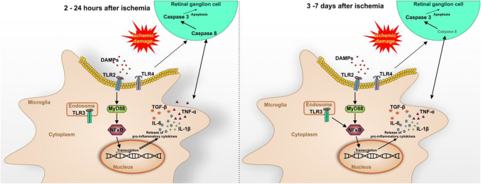
Mechanisms and progression of ischemic injuries in the retina are still incompletely clarified. Therefore, the time course of microglia activation as well as resulting cytokine expression and downstream signaling were investigated. Ischemia was induced in one eye by transiently elevated intraocular pressure (60 min) followed by reperfusion; the other eye served as a control. Eyes were processed for RT-qPCR and immunohistochemistry analyses at 2, 6, 12, and 24 h as well as at 3 and 7 days. Already 2 h after ischemia, more microglia/macrophages were in an active state in the ischemia group. This was accompanied by an upregulation of pro-inflammatory cytokines, like IL-1β, IL-6, TNFα, and TGFβ. Activation of TLR3, TLR2, and the adaptor molecule Myd88 was also observed after 2 h. NFκB revealed a wave-like activation pattern. In addition, an extrinsic caspase pathway activation was noted at early time points, while enhanced numbers of cleaved caspase 3+ cells could be observed in ischemic retinae throughout the study. Retinal ischemia induced an early and strong microglia/macrophage response as well as cytokine and apoptotic activation processes. Moreover, in early and late ischemic damaging processes, TLR expression and downstream signaling were involved, suggesting an involvement in neuronal death in ischemic retinae.
中文翻译:
视网膜缺血中的小胶质细胞激活触发细胞因子和 Toll 样受体反应。
视网膜缺血性损伤的机制和进展仍未完全阐明。因此,研究了小胶质细胞活化的时间过程以及由此产生的细胞因子表达和下游信号传导。通过短暂升高的眼压(60 分钟)然后再灌注在一只眼睛中诱导缺血;另一只眼睛作为对照。在 2、6、12 和 24 小时以及 3 和 7 天对眼睛进行 RT-qPCR 和免疫组织化学分析。缺血后 2 小时,缺血组中有更多的小胶质细胞/巨噬细胞处于活跃状态。这伴随着促炎细胞因子的上调,如 IL-1β、IL-6、TNFα 和 TGFβ。2 小时后还观察到 TLR3、TLR2 和衔接分子 Myd88 的激活。NFκB 揭示了一种波状激活模式。此外,在整个研究过程中,可以在缺血性视网膜中观察到+细胞。视网膜缺血诱导早期和强烈的小胶质细胞/巨噬细胞反应以及细胞因子和凋亡激活过程。此外,在早期和晚期缺血性损伤过程中,TLR 表达和下游信号传导参与,表明参与缺血性视网膜中的神经元死亡。


 京公网安备 11010802027423号