当前位置:
X-MOL 学术
›
Diam. Relat. Mater.
›
论文详情
Our official English website, www.x-mol.net, welcomes your feedback! (Note: you will need to create a separate account there.)
Electron Microscopy Study of Surface-Treated Carbon Fiber for Interface Modification in Composites
Diamond and Related Materials ( IF 4.1 ) Pub Date : 2020-11-01 , DOI: 10.1016/j.diamond.2020.108021 S.P. Sharma , J.M. Ting , R. Vilar
Diamond and Related Materials ( IF 4.1 ) Pub Date : 2020-11-01 , DOI: 10.1016/j.diamond.2020.108021 S.P. Sharma , J.M. Ting , R. Vilar
|
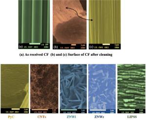
Abstract Relatively poor adhesion of carbon fibers with matrix leads to various serious concerns in high performance composites. Interface debonding during loading is the most vulnerable among them particularly when polymer matrix is used. This paper presents the results of electron microscopy investigation of four different techniques that are beneficial for improving interfacial bonding. These techniques were based on modifying the fiber surface by way of both; coating a film on the fiber and ablating a layer from it. The former employed growth of nanostructures of carbon and zinc oxide by thermal catalytic chemical vapor deposition and magnetron sputtering respectively and depositing a layer of pyrolytic carbon by CVD while the latter involved ablation of an outer layer of fiber using femtosecond laser treatment. Both the approaches relied on introducing mechanical anchoring on the fiber surface by micro roughening anticipating an improved fiber- matrix interfacial bonding. Carbon fiber surface prior and post treatment was investigated microscopically using FESEM and TEM. CNTs growth formed network like structure around the fiber, while PyC coating showed continuous layer around the fiber. HRTEM images of ZnO nanostructures; both nanowires and nanowalls possessed crystalline structure revealed by the presence of Moire fringes formed due to overlapping of two crystals with different orientation. Laser treated fiber surface showed formation of LIPSS with average periodicity of 147 ± 10 nm.
中文翻译:

用于复合材料界面改性的表面处理碳纤维的电子显微镜研究
摘要 碳纤维与基体的相对较差的粘附性导致高性能复合材料中的各种严重问题。加载过程中的界面脱粘是其中最脆弱的,尤其是在使用聚合物基质时。本文介绍了对改善界面结合有益的四种不同技术的电子显微镜研究结果。这些技术基于通过以下两种方式对纤维表面进行改性;在纤维上涂上一层薄膜并从它上面烧掉一层。前者分别通过热催化化学气相沉积和磁控溅射来生长碳和氧化锌的纳米结构,并通过 CVD 沉积一层热解碳,而后者涉及使用飞秒激光处理烧蚀外层纤维。这两种方法都依赖于通过微粗糙化在纤维表面引入机械锚定,以期改善纤维-基质界面结合。使用 FESEM 和 TEM 在显微镜下研究碳纤维表面处理前后。碳纳米管生长在纤维周围形成网络状结构,而 PyC 涂层在纤维周围显示连续层。ZnO纳米结构的HRTEM图像;纳米线和纳米壁都具有晶体结构,因为存在由于具有不同取向的两种晶体重叠而形成的莫尔条纹。激光处理的纤维表面显示形成平均周期为 147 ± 10 nm 的 LIPSS。使用 FESEM 和 TEM 在显微镜下研究碳纤维表面处理前后。碳纳米管生长在纤维周围形成网络状结构,而 PyC 涂层在纤维周围显示连续层。ZnO纳米结构的HRTEM图像;纳米线和纳米壁都具有晶体结构,因为存在由于具有不同取向的两种晶体重叠而形成的莫尔条纹。激光处理的纤维表面显示形成平均周期为 147 ± 10 nm 的 LIPSS。使用 FESEM 和 TEM 在显微镜下研究碳纤维表面处理前后。碳纳米管生长在纤维周围形成网络状结构,而 PyC 涂层在纤维周围显示连续层。ZnO纳米结构的HRTEM图像;纳米线和纳米壁都具有晶体结构,因为存在由于具有不同取向的两种晶体重叠而形成的莫尔条纹。激光处理的纤维表面显示形成平均周期为 147 ± 10 nm 的 LIPSS。纳米线和纳米壁都具有晶体结构,因为存在由于具有不同取向的两种晶体重叠而形成的莫尔条纹。激光处理的纤维表面显示形成平均周期为 147 ± 10 nm 的 LIPSS。纳米线和纳米壁都具有晶体结构,因为存在由于具有不同取向的两种晶体重叠而形成的莫尔条纹。激光处理的纤维表面显示形成平均周期为 147 ± 10 nm 的 LIPSS。
更新日期:2020-11-01
中文翻译:
用于复合材料界面改性的表面处理碳纤维的电子显微镜研究
摘要 碳纤维与基体的相对较差的粘附性导致高性能复合材料中的各种严重问题。加载过程中的界面脱粘是其中最脆弱的,尤其是在使用聚合物基质时。本文介绍了对改善界面结合有益的四种不同技术的电子显微镜研究结果。这些技术基于通过以下两种方式对纤维表面进行改性;在纤维上涂上一层薄膜并从它上面烧掉一层。前者分别通过热催化化学气相沉积和磁控溅射来生长碳和氧化锌的纳米结构,并通过 CVD 沉积一层热解碳,而后者涉及使用飞秒激光处理烧蚀外层纤维。这两种方法都依赖于通过微粗糙化在纤维表面引入机械锚定,以期改善纤维-基质界面结合。使用 FESEM 和 TEM 在显微镜下研究碳纤维表面处理前后。碳纳米管生长在纤维周围形成网络状结构,而 PyC 涂层在纤维周围显示连续层。ZnO纳米结构的HRTEM图像;纳米线和纳米壁都具有晶体结构,因为存在由于具有不同取向的两种晶体重叠而形成的莫尔条纹。激光处理的纤维表面显示形成平均周期为 147 ± 10 nm 的 LIPSS。使用 FESEM 和 TEM 在显微镜下研究碳纤维表面处理前后。碳纳米管生长在纤维周围形成网络状结构,而 PyC 涂层在纤维周围显示连续层。ZnO纳米结构的HRTEM图像;纳米线和纳米壁都具有晶体结构,因为存在由于具有不同取向的两种晶体重叠而形成的莫尔条纹。激光处理的纤维表面显示形成平均周期为 147 ± 10 nm 的 LIPSS。使用 FESEM 和 TEM 在显微镜下研究碳纤维表面处理前后。碳纳米管生长在纤维周围形成网络状结构,而 PyC 涂层在纤维周围显示连续层。ZnO纳米结构的HRTEM图像;纳米线和纳米壁都具有晶体结构,因为存在由于具有不同取向的两种晶体重叠而形成的莫尔条纹。激光处理的纤维表面显示形成平均周期为 147 ± 10 nm 的 LIPSS。纳米线和纳米壁都具有晶体结构,因为存在由于具有不同取向的两种晶体重叠而形成的莫尔条纹。激光处理的纤维表面显示形成平均周期为 147 ± 10 nm 的 LIPSS。纳米线和纳米壁都具有晶体结构,因为存在由于具有不同取向的两种晶体重叠而形成的莫尔条纹。激光处理的纤维表面显示形成平均周期为 147 ± 10 nm 的 LIPSS。


 京公网安备 11010802027423号