Colloids and Surfaces B: Biointerfaces ( IF 5.8 ) Pub Date : 2020-08-14 , DOI: 10.1016/j.colsurfb.2020.111320 Abner L Bogan 1 , Karen Fong 2 , Aljosa Trmcic 2 , Siyun Wang 2 , John M Frostad 3
|
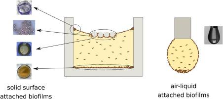
In food safety and food quality, biofilm research is of great importance for mitigating food-borne pathogens in food processing environments. To supplement the traditional staining techniques for biofilm characterization, we introduce several non-traditional imaging methods for detecting biofilm attachment to the solid–liquid and air–liquid interfaces. For strains of Pseudomonas aeruginosa (the positive control), Acinetobacter baumanii, Listeria monocytogenes and Salmonella enterica, the traditional crystal violet assay showed evidence of biofilm attachment to the well plate base as well as inferred the presence of an air–liquid biofilm attached on the upper well walls where the meniscus was present. However, air–liquid biofilms and solid-surface-attached biofilms were not detected for all of these strains using the non-traditional imaging methods. For L. monocytogenes, we were unable to detect biofilms at a particle-laden, air–liquid interface as evidenced through microscopy, which contradicts the meniscus staining test and suggests that the coffee-ring effect may lead to false positives when using meniscus staining. Furthermore, when L. monocytogenes was cultivated in a pendant droplet in air, only microbial sediment at the droplet apex was observed without any apparent bacterial colonization of the droplet surface. All other strains showed clear evidence of air–liquid biofilms at the air–liquid interface of a pendant droplet. To non-invasively detect if and when air–liquid pellicles form in a well plate, we also present a novel in situ reflection assay that demonstrates the capacity to do this quantitatively.
中文翻译:
评价非传统可视化方法以检测生物膜的表面附着。
在食品安全和食品质量方面,生物膜研究对于减轻食品加工环境中食源性病原体至关重要。为了补充用于生物膜表征的传统染色技术,我们引入了几种非传统的成像方法来检测生物膜在固-液和气-液界面上的附着。对于铜绿假单胞菌(阳性对照),鲍曼不动杆菌,单核细胞增生李斯特菌和肠炎沙门氏菌菌株传统的结晶紫测定法显示了生物膜附着在孔板底部的证据,并推断出弯液面所在的上部孔壁上存在气液生物膜的存在。但是,使用非传统成像方法未检测到所有这些菌株的气液生物膜和固体表面附着的生物膜。对于单核细胞增生李斯特氏菌,我们无法在显微镜观察到的充满颗粒的气液界面上检测到生物膜,这与半月板染色测试相矛盾,并表明使用半月板染色时咖啡环效应可能导致假阳性。此外,当单核细胞增生李斯特菌将其在空气中的悬滴中培养,仅观察到在滴顶点处的微生物沉积物,而在滴表面上没有任何明显的细菌定植。所有其他菌株均在悬滴的气液界面处显示出明显的气液生物膜证据。为了非侵入性地检测是否以及何时在气孔板上形成气膜薄膜,我们还提出了一种新颖的原位反射测定法,该测定法证明了进行定量分析的能力。


 京公网安备 11010802027423号