Our official English website, www.x-mol.net, welcomes your feedback! (Note: you will need to create a separate account there.)
Human Mesenchymal Stem Cell Derived Exosomes Enhance Cell‐Free Bone Regeneration by Altering Their miRNAs Profiles
Advanced Science ( IF 15.1 ) Pub Date : 2020-08-07 , DOI: 10.1002/advs.202001334 Mengmeng Zhai 1 , Ye Zhu 1 , Mingying Yang 2 , Chuanbin Mao 1, 3
Advanced Science ( IF 15.1 ) Pub Date : 2020-08-07 , DOI: 10.1002/advs.202001334 Mengmeng Zhai 1 , Ye Zhu 1 , Mingying Yang 2 , Chuanbin Mao 1, 3
Affiliation
|
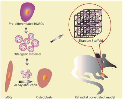
Implantation of stem cells for tissue regeneration faces significant challenges such as immune rejection and teratoma formation. Cell‐free tissue regeneration thus has a potential to avoid these problems. Stem cell derived exosomes do not cause immune rejection or generate malignant tumors. Here, exosomes that can induce osteogenic differentiation of human mesenchymal stem cells (hMSCs) are identified and used to decorate 3D‐printed titanium alloy scaffolds to achieve cell‐free bone regeneration. Specifically, the exosomes secreted by hMSCs osteogenically pre‐differentiated for different times are used to induce the osteogenesis of hMSCs. It is discovered that pre‐differentiation for 10 and 15 days leads to the production of osteogenic exosomes. The purified exosomes are then loaded into the scaffolds. It is found that the cell‐free exosome‐coated scaffolds regenerate bone tissue as efficiently as hMSC‐seeded exosome‐free scaffolds within 12 weeks. RNA‐sequencing suggests that the osteogenic exosomes induce the osteogenic differentiation by using their cargos, including upregulated osteogenic miRNAs (Hsa‐miR‐146a‐5p, Hsa‐miR‐503‐5p, Hsa‐miR‐483‐3p, and Hsa‐miR‐129‐5p) or downregulated anti‐osteogenic miRNAs (Hsa‐miR‐32‐5p, Hsa‐miR‐133a‐3p, and Hsa‐miR‐204‐5p), to activate the PI3K/Akt and MAPK signaling pathways. Consequently, identification of osteogenic exosomes secreted by pre‐differentiated stem cells and the use of them to replace stem cells represent a novel cell‐free bone regeneration strategy.
中文翻译:

人间充质干细胞衍生的外泌体通过改变 miRNA 谱来增强无细胞骨再生
植入干细胞进行组织再生面临着免疫排斥和畸胎瘤形成等重大挑战。因此,无细胞组织再生有可能避免这些问题。干细胞衍生的外泌体不会引起免疫排斥或产生恶性肿瘤。在此,鉴定出可诱导人间充质干细胞 (hMSC) 成骨分化的外泌体,并将其用于装饰 3D 打印的钛合金支架,以实现无细胞骨再生。具体来说,hMSCs成骨预分化不同时间后分泌的外泌体用于诱导hMSCs成骨。研究发现,预分化 10 天和 15 天会导致成骨外泌体的产生。然后将纯化的外泌体装载到支架中。研究发现,无细胞外泌体涂层支架在 12 周内再生骨组织的效率与接种 hMSC 的无外泌体支架一样有效。RNA测序表明,成骨外泌体通过使用其货物诱导成骨分化,包括上调的成骨miRNA(Hsa-miR-146a-5p、Hsa-miR-503-5p、Hsa-miR-483-3p和Hsa-miR) ‐129-5p) 或下调抗成骨 miRNA(Hsa-miR-32-5p、Hsa-miR-133a-3p 和 Hsa-miR-204-5p),以激活 PI3K/Akt 和 MAPK 信号通路。因此,鉴定预分化干细胞分泌的成骨外泌体并使用它们替代干细胞代表了一种新型的无细胞骨再生策略。
更新日期:2020-10-07
中文翻译:
人间充质干细胞衍生的外泌体通过改变 miRNA 谱来增强无细胞骨再生
植入干细胞进行组织再生面临着免疫排斥和畸胎瘤形成等重大挑战。因此,无细胞组织再生有可能避免这些问题。干细胞衍生的外泌体不会引起免疫排斥或产生恶性肿瘤。在此,鉴定出可诱导人间充质干细胞 (hMSC) 成骨分化的外泌体,并将其用于装饰 3D 打印的钛合金支架,以实现无细胞骨再生。具体来说,hMSCs成骨预分化不同时间后分泌的外泌体用于诱导hMSCs成骨。研究发现,预分化 10 天和 15 天会导致成骨外泌体的产生。然后将纯化的外泌体装载到支架中。研究发现,无细胞外泌体涂层支架在 12 周内再生骨组织的效率与接种 hMSC 的无外泌体支架一样有效。RNA测序表明,成骨外泌体通过使用其货物诱导成骨分化,包括上调的成骨miRNA(Hsa-miR-146a-5p、Hsa-miR-503-5p、Hsa-miR-483-3p和Hsa-miR) ‐129-5p) 或下调抗成骨 miRNA(Hsa-miR-32-5p、Hsa-miR-133a-3p 和 Hsa-miR-204-5p),以激活 PI3K/Akt 和 MAPK 信号通路。因此,鉴定预分化干细胞分泌的成骨外泌体并使用它们替代干细胞代表了一种新型的无细胞骨再生策略。


 京公网安备 11010802027423号