Journal of Neuroscience Methods ( IF 3 ) Pub Date : 2020-08-07 , DOI: 10.1016/j.jneumeth.2020.108903 Josefina Maranzano 1 , Mahsa Dadar 2 , Antony Bertrand-Grenier 3 , Eve-Marie Frigon 4 , Johanne Pellerin 4 , Sophie Plante 4 , Simon Duchesne 5 , Christine L Tardif 6 , Denis Boire 4 , Gilles Bronchti 4
|
Background
MRI-histology correlation studies of the ex vivo brain mostly employ fresh, extracted (ex situ) specimens, aldehyde fixed by immersion, which has several disadvantages for MRI scanning (e.g. deformation of the organ). A minority of studies are done ex vivo-in situ (unfixed brain), requiring an MRI scanner readily available within a few hours of the time of death.
New Method
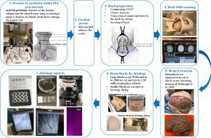
We propose a new technique, exploited by anatomists, for scanning the ex vivo brain: fixation by whole body perfusion, which implies fixation of the brain in situ. This allows scanning the brain surrounded by fluids, meninges, and skull, preserving the structural relationships of the brain in vivo.
To evaluate the proposed method, five heads perfused-fixed with a saturated sodium chloride solution were employed. Three sequences were acquired on a 1.5 T MRI scanner: T1weighted, T2weighted-FLAIR, and Gradient-echo. Histology analysis included immunofluorescence for myelin basic protein and neuronal nuclei.
Results
All MRIs were successfully processed through a validated pipeline used with in vivo MRIs. All cases exhibited positive antigenicity for myelin and neuronal nuclei.
Comparison with existing methods
All scans registered to a standard neuroanatomical template in pseudo-Talairach space more accurately than an ex vivo-ex situ scan.
The time interval to scan the ex vivo brain in situ was increased to at least 10 months.
Conclusions
MRI and histology study of the ex vivo-in situ brain fixed by perfusion is an alternative approach that has important procedural and practical advantages over the two standard methods to study the ex vivo brain.
中文翻译:
通过MRI和组织学研究人脑的一种新颖的离体原位方法。
背景
离体大脑的MRI-组织学相关性研究主要采用新鲜的,提取的(非原位)标本,通过浸没固定的醛,这对MRI扫描有几个缺点(例如器官变形)。少数研究是离体原位(未固定的大脑)进行的,需要在死亡后数小时内容易获得的MRI扫描仪。
新方法
我们提出了一种由解剖学家开发的用于扫描离体大脑的新技术:通过全身灌注进行固定,这意味着对大脑进行原位固定。这允许扫描被液体,脑膜和头骨包围的大脑,从而在体内保留大脑的结构关系。
为了评估所提出的方法,采用了五个用饱和氯化钠溶液灌注固定的探头。在1.5 T MRI扫描仪上采集了三个序列:T1加权,T2加权-FLAIR和梯度回波。组织学分析包括髓鞘碱性蛋白和神经元核的免疫荧光。
结果
所有MRI均通过与体内MRI一起使用的经过验证的管道成功处理。所有病例均显示出髓鞘和神经元核的阳性抗原性。
与现有方法的比较
与离体-异位扫描相比,所有扫描都更准确地注册到伪Talairach空间中的标准神经解剖模板。
原位扫描离体大脑的时间间隔增加到至少10个月。
结论
通过灌注固定离体原位脑的MRI和组织学研究是一种替代方法,它比研究离体脑的两种标准方法具有重要的程序和实践优势。


 京公网安备 11010802027423号