当前位置:
X-MOL 学术
›
Hum. Brain Mapp.
›
论文详情
Our official English website, www.x-mol.net, welcomes your feedback! (Note: you will need to create a separate account there.)
Improved Vim targeting for focused ultrasound ablation treatment of essential tremor: A probabilistic and patient-specific approach.
Human Brain Mapping ( IF 4.8 ) Pub Date : 2020-08-06 , DOI: 10.1002/hbm.25157 Jason H Su 1, 2 , Eun Young Choi 3 , Thomas Tourdias 4, 5 , Manojkumar Saranathan 6 , Casey H Halpern 3 , Jaimie M Henderson 3 , Kim Butts Pauly 1 , Pejman Ghanouni 1 , Brian K Rutt 1
Human Brain Mapping ( IF 4.8 ) Pub Date : 2020-08-06 , DOI: 10.1002/hbm.25157 Jason H Su 1, 2 , Eun Young Choi 3 , Thomas Tourdias 4, 5 , Manojkumar Saranathan 6 , Casey H Halpern 3 , Jaimie M Henderson 3 , Kim Butts Pauly 1 , Pejman Ghanouni 1 , Brian K Rutt 1
Affiliation
|
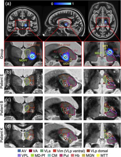
Magnetic resonance‐guided focused ultrasound (MRgFUS) ablation of the ventral intermediate (Vim) thalamic nucleus is an incisionless treatment for essential tremor (ET). The standard initial targeting method uses an approximate, atlas‐based stereotactic approach. We developed a new patient‐specific targeting method to identify an individual's Vim and the optimal MRgFUS target region therein for suppression of tremor. In this retrospective study of 14 ET patients treated with MRgFUS, we investigated the ability of WMnMPRAGE, a highly sensitive and robust sequence for imaging gray matter‐white matter contrast, to identify the Vim, FUS ablation, and a clinically efficacious region within the Vim in individual patients. We found that WMnMPRAGE can directly visualize the Vim in ET patients, segmenting this nucleus using manual or automated segmentation capabilities developed by our group. WMnMPRAGE also delineated the ablation's core and penumbra, and showed that all patients' ablation cores lay primarily within their Vim segmentations. We found no significant correlations between standard ablation features (e.g., ablation volume, Vim‐ablation overlap) and 1‐month post‐treatment clinical outcome. We then defined a group‐based probabilistic target, which was nonlinearly warped to individual brains; this target was located within the Vim for all patients. The overlaps between this target and patient ablation cores correlated significantly with 1‐month clinical outcome (r = −.57, p = .03), in contrast to the standard target (r = −.23, p = .44). We conclude that WMnMPRAGE is a highly sensitive sequence for segmenting Vim and ablation boundaries in individual patients, allowing us to find a novel tremor‐associated center within Vim and potentially improving MRgFUS treatment for ET.
中文翻译:

用于特发性震颤的聚焦超声消融治疗的改进 Vim 靶向:一种概率和患者特定的方法。
磁共振引导聚焦超声 (MRgFUS) 消融腹侧中间 (Vim) 丘脑核是特发性震颤 (ET) 的无切口治疗。标准的初始定位方法使用近似的、基于地图集的立体定向方法。我们开发了一种新的患者特异性靶向方法来识别个体的 Vim 和其中用于抑制震颤的最佳 MRgFUS 靶区。在这项对 14 名接受 MRgFUS 治疗的 ET 患者的回顾性研究中,我们研究了 WMnMPRAGE(一种用于灰质 - 白质对比成像的高度灵敏且稳健的序列)识别 Vim、FUS 消融和 Vim 内临床有效区域的能力在个别患者中。我们发现 WMnMPRAGE 可以直接可视化 ET 患者的 Vim,使用我们小组开发的手动或自动分割功能分割这个核心。WMnMPRAGE 还描绘了消融的核心和半影,并表明所有患者的消融核心主要位于他们的 Vim 分段内。我们发现标准消融特征(例如消融量、Vim 消融重叠)与治疗后 1 个月的临床结果之间没有显着相关性。然后我们定义了一个基于组的概率目标,该目标被非线性扭曲到个体大脑;该目标位于所有患者的 Vim 内。该目标和患者消融核心之间的重叠与 1 个月的临床结果显着相关(消融核心主要位于其 Vim 分段中。我们发现标准消融特征(例如消融量、Vim 消融重叠)与治疗后 1 个月的临床结果之间没有显着相关性。然后我们定义了一个基于组的概率目标,该目标被非线性扭曲到个体大脑;该目标位于所有患者的 Vim 内。该目标和患者消融核心之间的重叠与 1 个月的临床结果显着相关(消融核心主要位于其 Vim 分段中。我们发现标准消融特征(例如消融量、Vim 消融重叠)与治疗后 1 个月的临床结果之间没有显着相关性。然后我们定义了一个基于组的概率目标,该目标被非线性扭曲到个体大脑;该目标位于所有患者的 Vim 内。该目标和患者消融核心之间的重叠与 1 个月的临床结果显着相关(该目标位于所有患者的 Vim 内。该目标和患者消融核心之间的重叠与 1 个月的临床结果显着相关(该目标位于所有患者的 Vim 内。该目标和患者消融核心之间的重叠与 1 个月的临床结果显着相关(r = -.57, p = .03),与标准目标 ( r = -.23, p = .44)形成对比。我们得出结论,WMnMPRAGE 是一个高度敏感的序列,用于分割个体患者的 Vim 和消融边界,使我们能够在 Vim 中找到一个新的震颤相关中心,并有可能改善 ET 的 MRgFUS 治疗。
更新日期:2020-08-06
中文翻译:
用于特发性震颤的聚焦超声消融治疗的改进 Vim 靶向:一种概率和患者特定的方法。
磁共振引导聚焦超声 (MRgFUS) 消融腹侧中间 (Vim) 丘脑核是特发性震颤 (ET) 的无切口治疗。标准的初始定位方法使用近似的、基于地图集的立体定向方法。我们开发了一种新的患者特异性靶向方法来识别个体的 Vim 和其中用于抑制震颤的最佳 MRgFUS 靶区。在这项对 14 名接受 MRgFUS 治疗的 ET 患者的回顾性研究中,我们研究了 WMnMPRAGE(一种用于灰质 - 白质对比成像的高度灵敏且稳健的序列)识别 Vim、FUS 消融和 Vim 内临床有效区域的能力在个别患者中。我们发现 WMnMPRAGE 可以直接可视化 ET 患者的 Vim,使用我们小组开发的手动或自动分割功能分割这个核心。WMnMPRAGE 还描绘了消融的核心和半影,并表明所有患者的消融核心主要位于他们的 Vim 分段内。我们发现标准消融特征(例如消融量、Vim 消融重叠)与治疗后 1 个月的临床结果之间没有显着相关性。然后我们定义了一个基于组的概率目标,该目标被非线性扭曲到个体大脑;该目标位于所有患者的 Vim 内。该目标和患者消融核心之间的重叠与 1 个月的临床结果显着相关(消融核心主要位于其 Vim 分段中。我们发现标准消融特征(例如消融量、Vim 消融重叠)与治疗后 1 个月的临床结果之间没有显着相关性。然后我们定义了一个基于组的概率目标,该目标被非线性扭曲到个体大脑;该目标位于所有患者的 Vim 内。该目标和患者消融核心之间的重叠与 1 个月的临床结果显着相关(消融核心主要位于其 Vim 分段中。我们发现标准消融特征(例如消融量、Vim 消融重叠)与治疗后 1 个月的临床结果之间没有显着相关性。然后我们定义了一个基于组的概率目标,该目标被非线性扭曲到个体大脑;该目标位于所有患者的 Vim 内。该目标和患者消融核心之间的重叠与 1 个月的临床结果显着相关(该目标位于所有患者的 Vim 内。该目标和患者消融核心之间的重叠与 1 个月的临床结果显着相关(该目标位于所有患者的 Vim 内。该目标和患者消融核心之间的重叠与 1 个月的临床结果显着相关(r = -.57, p = .03),与标准目标 ( r = -.23, p = .44)形成对比。我们得出结论,WMnMPRAGE 是一个高度敏感的序列,用于分割个体患者的 Vim 和消融边界,使我们能够在 Vim 中找到一个新的震颤相关中心,并有可能改善 ET 的 MRgFUS 治疗。


 京公网安备 11010802027423号