当前位置:
X-MOL 学术
›
Soft Matter
›
论文详情
Our official English website, www.x-mol.net, welcomes your feedback! (Note: you will need to create a separate account there.)
Peak force visible microscopy.
Soft Matter ( IF 3.4 ) Pub Date : 2020-08-03 , DOI: 10.1039/d0sm01104e Haomin Wang 1 , Le Wang 1 , Yuequn Shang 2 , Sajedehalsadat Yazdanparast Tafti 3 , Wenpeng Cao 3 , Zhijun Ning 2 , X Frank Zhang 3 , Xiaoji G Xu 1
Soft Matter ( IF 3.4 ) Pub Date : 2020-08-03 , DOI: 10.1039/d0sm01104e Haomin Wang 1 , Le Wang 1 , Yuequn Shang 2 , Sajedehalsadat Yazdanparast Tafti 3 , Wenpeng Cao 3 , Zhijun Ning 2 , X Frank Zhang 3 , Xiaoji G Xu 1
Affiliation
|
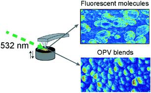
The optical responses of molecules and materials provide a basis for chemical measurement and imaging. The optical diffraction limit in conventional light microscopy is exceeded by mechanically probing optical absorption through the photothermal effect with atomic force microscopy (AFM). However, the spatial resolution of AFM-based photothermal optical microscopy is still limited, and the sample surface is prone to damage from scratching due to tip contact, particularly for measurements on soft matter. In this article, we develop peak force visible (PF-vis) microscopy for the measurement of visible optical absorption of soft matter. The spatial resolution of PF-vis microscopy is demonstrated to be 3 nm on green fluorescent protein-labeled virus-like particles, and the imaging sensitivity may approach a single protein molecule. On organic photovoltaic polymers, the spatial distribution of the optical absorption probed by PF-vis microscopy is found to be dependent on the diffusion ranges of excitons in the donor domain. Through finite element modeling and data analysis, the exciton diffusion range of organic photovoltaics can be directly extracted from PF-vis images, saving the need for complex and delicate sample preparations. PF-vis microscopy will enable high-resolution nano-imaging based on light absorption of fluorophores and chromophores, as well as deciphering the correlation between the spatial distribution of photothermal signals and underlying photophysical parameters at the tens of nanometer scale.
中文翻译:

峰值力可见显微镜。
分子和材料的光学响应为化学测量和成像提供了基础。通过利用原子力显微镜(AFM)的光热效应通过机械方式探测光吸收,超出了常规光学显微镜的光学衍射极限。但是,基于AFM的光热光学显微镜的空间分辨率仍然受到限制,并且由于针尖的接触,特别是对于软物质的测量,样品表面容易受到刮擦的损害。在本文中,我们开发了峰值力可见光(PF-vis)显微镜,用于测量软物质的可见光吸收。PF-vis显微镜的空间分辨率在绿色荧光蛋白标记的病毒样颗粒上显示为3 nm,并且成像灵敏度可能接近单个蛋白分子。在有机光伏聚合物上,发现通过PF-vis显微镜探测的光吸收的空间分布取决于激子在供体域中的扩散范围。通过有限元建模和数据分析,可以直接从PF-vis图像中提取有机光伏激子的扩散范围,从而无需进行复杂而精细的样品制备。PF-vis显微镜将实现基于荧光团和生色团的光吸收的高分辨率纳米成像,以及在数十纳米尺度上解读光热信号的空间分布与潜在的光物理参数之间的相关性。发现通过PF-vis显微镜探测的光吸收的空间分布取决于激子在供体域中的扩散范围。通过有限元建模和数据分析,可以直接从PF-vis图像中提取有机光伏激子的扩散范围,从而无需进行复杂而精细的样品制备。PF-vis显微镜将实现基于荧光团和生色团的光吸收的高分辨率纳米成像,以及在数十纳米尺度上解读光热信号的空间分布与潜在的光物理参数之间的相关性。发现通过PF-vis显微镜探测的光吸收的空间分布取决于激子在供体域中的扩散范围。通过有限元建模和数据分析,可以直接从PF-vis图像中提取有机光伏激子的扩散范围,从而节省了复杂而精细的样品制备工作。PF-vis显微镜将实现基于荧光团和生色团的光吸收的高分辨率纳米成像,以及在数十纳米尺度上解读光热信号的空间分布与潜在的光物理参数之间的相关性。节省了复杂而微妙的样品制备需求。PF-vis显微镜将实现基于荧光团和生色团的光吸收的高分辨率纳米成像,以及在数十纳米尺度上解读光热信号的空间分布与潜在的光物理参数之间的相关性。节省了复杂而微妙的样品制备需求。PF-vis显微镜将实现基于荧光团和发色团的光吸收的高分辨率纳米成像,以及在数十纳米尺度上解读光热信号的空间分布与潜在的光物理参数之间的相关性。
更新日期:2020-09-23
中文翻译:
峰值力可见显微镜。
分子和材料的光学响应为化学测量和成像提供了基础。通过利用原子力显微镜(AFM)的光热效应通过机械方式探测光吸收,超出了常规光学显微镜的光学衍射极限。但是,基于AFM的光热光学显微镜的空间分辨率仍然受到限制,并且由于针尖的接触,特别是对于软物质的测量,样品表面容易受到刮擦的损害。在本文中,我们开发了峰值力可见光(PF-vis)显微镜,用于测量软物质的可见光吸收。PF-vis显微镜的空间分辨率在绿色荧光蛋白标记的病毒样颗粒上显示为3 nm,并且成像灵敏度可能接近单个蛋白分子。在有机光伏聚合物上,发现通过PF-vis显微镜探测的光吸收的空间分布取决于激子在供体域中的扩散范围。通过有限元建模和数据分析,可以直接从PF-vis图像中提取有机光伏激子的扩散范围,从而无需进行复杂而精细的样品制备。PF-vis显微镜将实现基于荧光团和生色团的光吸收的高分辨率纳米成像,以及在数十纳米尺度上解读光热信号的空间分布与潜在的光物理参数之间的相关性。发现通过PF-vis显微镜探测的光吸收的空间分布取决于激子在供体域中的扩散范围。通过有限元建模和数据分析,可以直接从PF-vis图像中提取有机光伏激子的扩散范围,从而无需进行复杂而精细的样品制备。PF-vis显微镜将实现基于荧光团和生色团的光吸收的高分辨率纳米成像,以及在数十纳米尺度上解读光热信号的空间分布与潜在的光物理参数之间的相关性。发现通过PF-vis显微镜探测的光吸收的空间分布取决于激子在供体域中的扩散范围。通过有限元建模和数据分析,可以直接从PF-vis图像中提取有机光伏激子的扩散范围,从而节省了复杂而精细的样品制备工作。PF-vis显微镜将实现基于荧光团和生色团的光吸收的高分辨率纳米成像,以及在数十纳米尺度上解读光热信号的空间分布与潜在的光物理参数之间的相关性。节省了复杂而微妙的样品制备需求。PF-vis显微镜将实现基于荧光团和生色团的光吸收的高分辨率纳米成像,以及在数十纳米尺度上解读光热信号的空间分布与潜在的光物理参数之间的相关性。节省了复杂而微妙的样品制备需求。PF-vis显微镜将实现基于荧光团和发色团的光吸收的高分辨率纳米成像,以及在数十纳米尺度上解读光热信号的空间分布与潜在的光物理参数之间的相关性。


 京公网安备 11010802027423号