当前位置:
X-MOL 学术
›
J. Mater. Chem. B
›
论文详情
Our official English website, www.x-mol.net, welcomes your feedback! (Note: you will need to create a separate account there.)
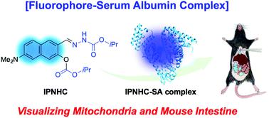
Visualizing mitochondria and mouse intestine with a fluorescent complex of a naphthalene-based dipolar dye and serum albumin.
Journal of Materials Chemistry B ( IF 7 ) Pub Date : 2020-07-22 , DOI: 10.1039/d0tb01314e Jong Min An 1 , Heejo Moon , Yejin Kim , Sangrim Kang , Youngseo Kim , Yuna Jung , Sungnam Park , Peter Verwilst , B Moon Kim , Jae Seung Kang , Dokyoung Kim
Journal of Materials Chemistry B ( IF 7 ) Pub Date : 2020-07-22 , DOI: 10.1039/d0tb01314e Jong Min An 1 , Heejo Moon , Yejin Kim , Sangrim Kang , Youngseo Kim , Yuna Jung , Sungnam Park , Peter Verwilst , B Moon Kim , Jae Seung Kang , Dokyoung Kim
Affiliation
|
We have explored a new research field of fluorophores through the manipulation of fluorophore-binding proteins. The development of a new imaging agent for tracing a specific organelle or a particular site within a living organism has been of great interest in the field of basic science as well as translational medicine. In this work and for the first time, we will disclose a new naphthalene-based dipolar dye and its complex, with serum albumin (SA), and show their applicability for the selective imaging of mitochondria in cells and the intestine in a mouse. The SA-binding dipolar dye, IPNHC, was synthesized straightforwardly, and we identified its photophysical properties and binding mode with SA. IPNHC–SA complex showed a bright emission in the blue wavelength range with a high quantum yield and stability. In the fluorescence imaging study, bright fluorescence images of mouse intestines were observed under a UV light, as well as two-photon (TP) deep tissue imaging after intravenous injection of IPNHC and IPNHC–SA complex. The present findings hold great promise for the application of the fluorescent complex for use in the tracing and tracking of intestine-related diseases at clinical sites.
中文翻译:

用基于萘的偶极染料和血清白蛋白的荧光复合物可视化线粒体和小鼠肠道。
我们已经通过操纵荧光团结合蛋白探索了一个新的荧光团研究领域。在基础科学和转化医学领域中,对于追踪生物体内特定细胞器或特定部位的新型成像剂的开发一直很感兴趣。在这项工作中,并且是第一次,我们将公开一种新型的萘基偶极染料及其配合物,与血清白蛋白(SA)结合使用,并展示它们在小鼠和细胞中线粒体选择性成像中的适用性。结合SA的偶极染料IPNHC可以直接合成,我们确定了它的光物理性质和与SA的结合方式。IPNHC–SA络合物在蓝色波长范围内显示出明亮的发射,具有很高的量子产率和稳定性。在荧光成像研究中 在静脉内注射IPNHC和IPNHC–SA复合物后,在紫外线和双光子(TP)深组织成像下观察到小鼠肠的明亮荧光图像。本发现为荧光复合物在临床部位肠道相关疾病的追踪中的应用提供了广阔的前景。
更新日期:2020-09-02
中文翻译:
用基于萘的偶极染料和血清白蛋白的荧光复合物可视化线粒体和小鼠肠道。
我们已经通过操纵荧光团结合蛋白探索了一个新的荧光团研究领域。在基础科学和转化医学领域中,对于追踪生物体内特定细胞器或特定部位的新型成像剂的开发一直很感兴趣。在这项工作中,并且是第一次,我们将公开一种新型的萘基偶极染料及其配合物,与血清白蛋白(SA)结合使用,并展示它们在小鼠和细胞中线粒体选择性成像中的适用性。结合SA的偶极染料IPNHC可以直接合成,我们确定了它的光物理性质和与SA的结合方式。IPNHC–SA络合物在蓝色波长范围内显示出明亮的发射,具有很高的量子产率和稳定性。在荧光成像研究中 在静脉内注射IPNHC和IPNHC–SA复合物后,在紫外线和双光子(TP)深组织成像下观察到小鼠肠的明亮荧光图像。本发现为荧光复合物在临床部位肠道相关疾病的追踪中的应用提供了广阔的前景。


 京公网安备 11010802027423号