Tissue & Cell ( IF 2.6 ) Pub Date : 2020-07-13 , DOI: 10.1016/j.tice.2020.101409 Hichem Kacem 1 , Jordi Miquel 2
|
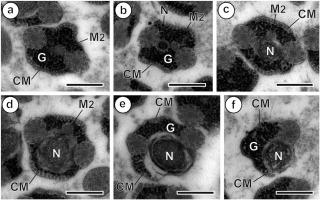
The ultrastructural characteristics of the mature spermatozoon of Holorchis pycnoporus (Digenea, Lepocreadioidea, Aephnidiogenidae) are described by means of transmission electron microscopy (TEM). Live worms were collected from the digestive tract of the Striped seabream Lithognathus mormyrus (Teleostei, Sparidae), off the Gulf of Gabès at La Chebba (Tunisia). The ultrastructural study reveals that the male gamete of H. pycnoporus is a filiform cell tapered at both extremities and exhibiting the type III of the digenean spermatozoon proposed by Bakhoum et al. (2017a), characterized by the presence of (1) two axonemes with the 9 + ‘1’ pattern of the Trepaxonemata, (2) external ornamentation of the plasma membrane located in a posterior part of the anterior region of the spermatozoon and associated with cortical microtubules, (3) two bundles of parallel cortical microtubules with maximum number located in the middle part of the spermatozoon, and (4) generally two mitochondria. Moreover, H. pycnoporus shares a set of ultrastructural characteristics with the studied Aephnidiogenidae such as: (1) two 9+'1' axonemes of different lengths, (2) an anterior electron-dense material, (3) mitochondrion/a, (4) an external ornamentation of the plasma membrane associated with cortical microtubules, and (5) two bundles of parallel cortical microtubules with their maximum number (around 24 microtubules) located in the middle or posterior part of the spermatozoon. In the Aephnidiogenidae, the mature spermatozoon exhibits a similar ultrastructural pattern. Some differences are observed, particularly the location of maximum number of cortical microtubules and the number of mitochondria. The presence of the anterolateral electron-dense material is the major particularity in species belonging to the Lepocreadioidea. This anterior dense material could be a synapomorphy for the superfamily and an ultrastructural argument supporting the monophyletic status of the Lepocreadioidea (Bray and Cribb, 2012).
中文翻译:
Lepocreadioidea 中的精子学特征,以及关于 Holorchis pycnoporus(Aephnidiogenidae)的第一个数据,这是来自加贝斯湾(突尼斯)的条纹鲷鱼 Lithognathus mormyrus(Sparidae)的寄生虫。
Holorchis pycnoporus (Digenea, Lepocreadioidea, Aephnidiogenidae)成熟精子的超微结构特征通过透射电子显微镜 (TEM) 进行描述。从La Chebba(突尼斯)的 Gabès 湾附近的条纹鲷鱼Lithognathus mormyrus(Teleostei,Sparidae)的消化道收集活蠕虫。超微结构研究表明H. pycnoporus的雄配子是一种在两端逐渐变细的丝状细胞,表现出 Bakhoum 等人提出的 digenean 精子的 III 型。(2017a),其特征在于存在 (1) 两个轴丝,具有 9 + '1' 模式的 Trepaxonemata,(2) 位于精子前部区域后部的质膜的外部装饰,并与皮层微管,(3)两束平行的皮层微管,数量最多,位于精子的中部,(4)一般有两个线粒体。此外,H. pycnoporus与所研究的 Aephnidiogenidae 共享一组超微结构特征,例如:(1) 两个不同长度的 9+'1' 轴丝,(2) 前电子致密材料,(3) 线粒体/a,(4) 外部装饰与皮质微管相关的质膜,以及 (5) 两束平行的皮质微管,它们的最大数量(约 24 个微管)位于精子的中部或后部。在 Aephnidiogenidae 中,成熟的精子表现出类似的超微结构模式。观察到一些差异,特别是皮质微管的最大数量和线粒体数量的位置。前外侧电子致密物质的存在是属于 Lepocreadioidea 的物种的主要特征。


 京公网安备 11010802027423号