当前位置:
X-MOL 学术
›
J. Raman Spectrosc.
›
论文详情
Our official English website, www.x-mol.net, welcomes your feedback! (Note: you will need to create a separate account there.)
Single‐beam optogenetic multimodal χ(3)/χ(5) nonlinear microscopy and brain imaging
Journal of Raman Spectroscopy ( IF 2.5 ) Pub Date : 2020-07-12 , DOI: 10.1002/jrs.5933 Aleksandr A. Lanin 1, 2, 3 , Artem S. Chebotarev 1 , Matvey S. Pochechuev 1 , Ilya V. Kelmanson 4, 5 , Daria A. Kotova 4 , Dmitry S. Bilan 4, 5 , Anatoly A. Ivanov 1, 6 , Anastasiya S. Panova 4, 5 , Viktor S. Tarabykin 7 , Andrei B. Fedotov 1, 2, 3, 8 , Vsevolod V. Belousov 4, 5, 9 , Aleksei M. Zheltikov 1, 2, 3, 10
Journal of Raman Spectroscopy ( IF 2.5 ) Pub Date : 2020-07-12 , DOI: 10.1002/jrs.5933 Aleksandr A. Lanin 1, 2, 3 , Artem S. Chebotarev 1 , Matvey S. Pochechuev 1 , Ilya V. Kelmanson 4, 5 , Daria A. Kotova 4 , Dmitry S. Bilan 4, 5 , Anatoly A. Ivanov 1, 6 , Anastasiya S. Panova 4, 5 , Viktor S. Tarabykin 7 , Andrei B. Fedotov 1, 2, 3, 8 , Vsevolod V. Belousov 4, 5, 9 , Aleksei M. Zheltikov 1, 2, 3, 10
Affiliation
|
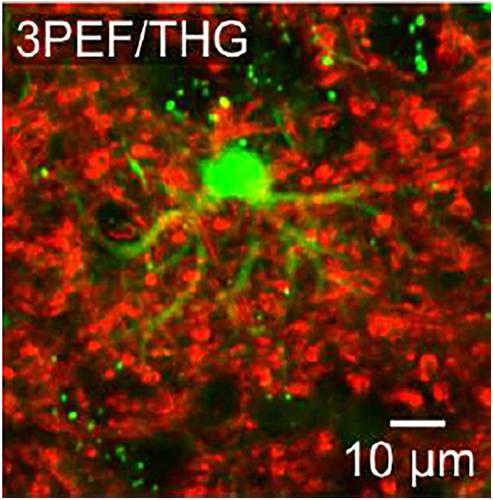
We demonstrate single‐beam optogenetic multimodal nonlinear‐optical microscopy that combines third‐harmonic generation (THG) and three‐photon‐excited fluorescence (3PEF) – two nonlinear‐optical processes related to the third‐ and fifth‐order susceptibilities, χ(3) and χ(5). A carefully tailored unamplified short‐pulse output of mode‐locked solid‐state lasers is shown to provide an ample parameter space for the optimization of such a single‐beam multimodal microscopy, enabling subcellular‐resolution, cell‐specific imaging using genetically encoded fluorescent‐protein‐based reporters in a vast variety of biological systems and settings, ranging from HeLa to brain cells. Experiments on brain slices and cell cultures presented in this paper demonstrate the potential of short‐pulse 3PEF/THG microscopy for a subcellular‐resolution, high‐contrast imaging of HeLa‐line cancer‐cell derivatives, as well as mitochondrial and somatic intracellular structures within deep‐brain neurons and astrocytes. As a step beyond the state of the art in optical brain imaging, single‐beam subcellular‐resolution, cell‐specific optogenetic 3PEF/THG imaging of fundamental functional brain units is experimentally demonstrated. One fundamental question that this work brings up for the future nonlinear absorption and Raman/hyper‐Raman studies is whether the Herzberg–Teller corrections, needed for an accurate description of two‐photon absorption in fluorescent proteins (FPs), would be also sufficient for an adequate treatment of higher‐n n‐photon absorption in FP‐based systems or models that include other quantum pathways would be necessary for the analysis of FP agents for higher‐n nonlinear microscopy.
中文翻译:

单光束光遗传多峰χ(3)/χ(5)非线性显微镜和脑成像
我们展示了结合了三次谐波产生(THG)和三光子激发荧光(3PEF)的单光束光遗传多峰非线性光学显微镜-与第三和第五级磁化率χ (3 )和χ (5)。精心定制的锁模固态激光器的短脉冲输出显示出可为此类单光束多峰显微镜的优化提供充足的参数空间,从而能够使用遗传编码的荧光光谱仪进行亚细胞分辨率的细胞特异性成像。从HeLa到脑细胞的各种生物系统和环境中的蛋白质报告基因。本文介绍的脑切片和细胞培养物实验证明了短脉冲3PEF / THG显微镜对于HeLa系癌细胞衍生物的亚细胞分辨率,高对比度成像以及其中的线粒体和体细胞内结构的潜力深脑神经元和星形胶质细胞。作为光学脑成像技术的最新发展,单光束亚细胞分辨率,基本的脑功能单元的细胞特异性光遗传学3PEF / THG成像已通过实验证明。这项工作为未来的非线性吸收和拉曼/超拉曼研究提出了一个基本问题,即准确描述荧光蛋白(FPs)中的双光子吸收所需的Herzberg-Teller校正是否也足以满足要求。对较高基于FP的系统或模型(包括其他量子路径)中的nn光子吸收对于高n非线性显微镜的FP剂分析而言是必要的。
更新日期:2020-07-12
中文翻译:
单光束光遗传多峰χ(3)/χ(5)非线性显微镜和脑成像
我们展示了结合了三次谐波产生(THG)和三光子激发荧光(3PEF)的单光束光遗传多峰非线性光学显微镜-与第三和第五级磁化率χ (3 )和χ (5)。精心定制的锁模固态激光器的短脉冲输出显示出可为此类单光束多峰显微镜的优化提供充足的参数空间,从而能够使用遗传编码的荧光光谱仪进行亚细胞分辨率的细胞特异性成像。从HeLa到脑细胞的各种生物系统和环境中的蛋白质报告基因。本文介绍的脑切片和细胞培养物实验证明了短脉冲3PEF / THG显微镜对于HeLa系癌细胞衍生物的亚细胞分辨率,高对比度成像以及其中的线粒体和体细胞内结构的潜力深脑神经元和星形胶质细胞。作为光学脑成像技术的最新发展,单光束亚细胞分辨率,基本的脑功能单元的细胞特异性光遗传学3PEF / THG成像已通过实验证明。这项工作为未来的非线性吸收和拉曼/超拉曼研究提出了一个基本问题,即准确描述荧光蛋白(FPs)中的双光子吸收所需的Herzberg-Teller校正是否也足以满足要求。对较高基于FP的系统或模型(包括其他量子路径)中的nn光子吸收对于高n非线性显微镜的FP剂分析而言是必要的。


 京公网安备 11010802027423号