Neuroscience ( IF 3.3 ) Pub Date : 2020-07-09 , DOI: 10.1016/j.neuroscience.2020.07.006 Rohan V Gupta 1 , Angel J Santiago-Lopez 2 , Ken Berglund 3 , Robert E Gross 4 , Claire-Anne N Gutekunst 1
|
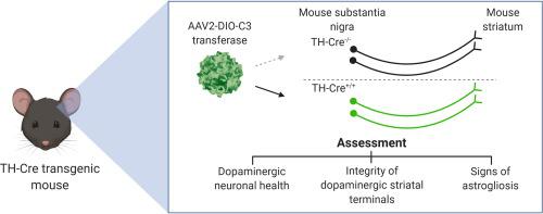
The bacterial exoenzyme C3 transferase (C3) irreversibly inhibits RhoA GTPase leading to stimulation of axonal outgrowth in injured neurons. C3 has been used successfully in models of neurotrauma and shows promise as an option to support cell survival and axonal growth of dopaminergic (DA) neurons in Parkinson’s disease (PD) cell therapy. Whether the continuous expression of C3 in DA neurons is well-tolerated is unknown. To assess the potential neurotoxicity of sustained expression of C3 in DA neurons, we generated Cre recombinase-dependent adeno-associated viral vectors (AAV) for targeted C3 delivery to DA neurons of the mouse substantia nigra pars compacta (SNc). The effect of continuous expression of C3 on DA neurons was assessed by immunohistochemistry and compared to that of Enhanced Yellow Fluorescent Protein (EYFP) as negative controls. We did not find significant reduction of tyrosine hydroxylase (TH) expression levels nor the presence of cleaved activated caspase 3. Astrocytic activation as determined by GFAP expression was comparable to EYFP controls. To evaluate the impact of C3 expression on striatal terminals of the nigrostriatal pathway, we compared the rotational behavior of wildtype mice injected unilaterally with either C3 or 6-hydroxydopamine (6-OHDA). Mice injected with C3 exhibited similar ipsiversive rotations to the site of injection in comparison to control mice injected with EYFP and significantly fewer ipsiversive rotations compared to 6-OHDA lesioned mice. Non-significant difference between C3 and EYFP controls in behavioral and histological analyses demonstrate that transduced DA neurons express C3 continuously without apparent adverse effects, supporting the use of C3 in efficacy studies targeting DA neurons.
中文翻译:
C3 转移酶连续表达后细胞死亡和黑质纹状体通路完整性的体内评估。
细菌外酶 C3 转移酶 (C3) 不可逆地抑制 RhoA GTPase,从而刺激受损神经元的轴突生长。C3 已成功用于神经创伤模型,并有望成为支持帕金森病 (PD) 细胞治疗中多巴胺能 (DA) 神经元的细胞存活和轴突生长的一种选择。DA 神经元中 C3 的持续表达是否被良好耐受尚不清楚。为了评估 C3 在 DA 神经元中持续表达的潜在神经毒性,我们生成了 Cre 重组酶依赖性腺相关病毒载体 (AAV),用于将 C3 靶向递送至小鼠黑质致密部 (SNc) 的 DA 神经元。通过免疫组织化学评估 C3 连续表达对 DA 神经元的影响,并与作为阴性对照的增强型黄色荧光蛋白 (EYFP) 进行比较。我们没有发现酪氨酸羟化酶 (TH) 表达水平显着降低,也没有发现切割的活化 caspase 3 的存在。由 GFAP 表达确定的星形胶质细胞活化与 EYFP 对照相当。为了评估 C3 表达对黑质纹状体通路纹状体末端的影响,我们比较了单侧注射 C3 或 6-羟基多巴胺 (6-OHDA) 的野生型小鼠的旋转行为。与注射 EYFP 的对照小鼠相比,注射 C3 的小鼠表现出与注射部位相似的同向旋转,并且与 6-OHDA 损伤小鼠相比显着更少的同向旋转。在行为和组织学分析中,C3 和 EYFP 对照之间的非显着差异表明,转导的 DA 神经元持续表达 C3 而没有明显的副作用,


 京公网安备 11010802027423号