当前位置:
X-MOL 学术
›
Cytom. Part A
›
论文详情
Our official English website, www.x-mol.net, welcomes your feedback! (Note: you will need to create a separate account there.)
Using Cytometry for Investigation of Purinergic Signaling in Tumor-Associated Macrophages.
Cytometry Part A ( IF 3.7 ) Pub Date : 2020-07-07 , DOI: 10.1002/cyto.a.24035 Vanessa F Arnaud-Sampaio 1 , Izadora L A Rabelo 1 , Carolina A Bento 1 , Talita Glaser 1 , Jean Bezerra 1 , Robson Coutinho-Silva 2 , Henning Ulrich 1 , Claudiana Lameu 1
Cytometry Part A ( IF 3.7 ) Pub Date : 2020-07-07 , DOI: 10.1002/cyto.a.24035 Vanessa F Arnaud-Sampaio 1 , Izadora L A Rabelo 1 , Carolina A Bento 1 , Talita Glaser 1 , Jean Bezerra 1 , Robson Coutinho-Silva 2 , Henning Ulrich 1 , Claudiana Lameu 1
Affiliation
|
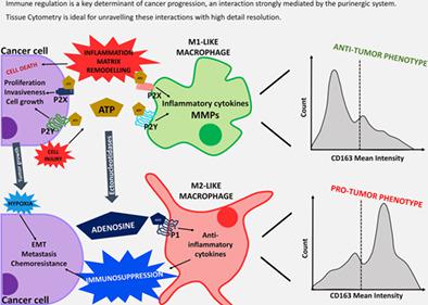
Tumor‐associated macrophages are widely recognized for their importance in guiding pro‐tumoral or antitumoral responses. Mediating inflammation or immunosuppression, these cells support many key events in cancer progression: cell growth, chemotaxis, invasiveness, angiogenesis and cell death. The communication between cells in the tumor microenvironment strongly relies on the secretion and recognition of several molecules, including damage‐associated molecular patterns (DAMPs), such as adenosine triphosphate (ATP). Extracellular ATP (eATP) and its degradation products act as signaling molecules and have extensively described roles in immune response and inflammation, as well as in cancer biology. These multiple functions highlight the purinergic system as a promising target to investigate the interplay between macrophages and cancer cells. Here, we reviewed purinergic signaling pathways connecting cancer cells and macrophages, a yet poorly investigated field. Finally, we present a new tool for the characterization of macrophage phenotype within the tumor. Image cytometry emerges as a cutting‐edge tool, capable of providing a broad set of information on cell morphology, expression of specific markers, and its cellular or subcellular localization, preserving cell–cell interactions within the tumor section and providing high statistical strength in small‐sized experiments. Thus, image cytometry allows deeper investigation of tumor heterogeneity and interactions between these cells. © 2020 International Society for Advancement of Cytometry
中文翻译:

使用细胞计数法研究肿瘤相关巨噬细胞中的嘌呤能信号传导。
肿瘤相关巨噬细胞因其在指导促肿瘤或抗肿瘤反应方面的重要性而被广泛认可。这些细胞介导炎症或免疫抑制,支持癌症进展中的许多关键事件:细胞生长、趋化性、侵袭性、血管生成和细胞死亡。肿瘤微环境中细胞之间的通讯强烈依赖于几种分子的分泌和识别,包括损伤相关分子模式 (DAMP),如三磷酸腺苷 (ATP)。细胞外 ATP (eATP) 及其降解产物充当信号分子,并广泛描述了在免疫反应和炎症以及癌症生物学中的作用。这些多重功能突出了嘌呤能系统作为研究巨噬细胞和癌细胞之间相互作用的有希望的目标。这里,我们回顾了连接癌细胞和巨噬细胞的嘌呤能信号通路,这是一个尚未得到充分研究的领域。最后,我们提出了一种用于表征肿瘤内巨噬细胞表型的新工具。图像细胞术作为一种尖端工具出现,能够提供关于细胞形态、特定标志物的表达及其细胞或亚细胞定位的广泛信息,保留肿瘤切片内的细胞-细胞相互作用,并在小细胞中提供高统计强度。规模的实验。因此,图像细胞术可以更深入地研究肿瘤异质性和这些细胞之间的相互作用。© 2020 国际细胞计量学促进会 我们提出了一种表征肿瘤内巨噬细胞表型的新工具。图像细胞术作为一种尖端工具出现,能够提供关于细胞形态、特定标志物的表达及其细胞或亚细胞定位的广泛信息,保留肿瘤切片内的细胞-细胞相互作用,并在小细胞中提供高统计强度。规模的实验。因此,图像细胞术可以更深入地研究肿瘤异质性和这些细胞之间的相互作用。© 2020 国际细胞计量学促进会 我们提出了一种表征肿瘤内巨噬细胞表型的新工具。图像细胞术作为一种尖端工具出现,能够提供关于细胞形态、特定标志物的表达及其细胞或亚细胞定位的广泛信息,保留肿瘤切片内的细胞-细胞相互作用,并在小细胞中提供高统计强度。规模的实验。因此,图像细胞术可以更深入地研究肿瘤异质性和这些细胞之间的相互作用。© 2020 国际细胞计量学促进会 保留肿瘤切片内的细胞间相互作用,并在小规模实验中提供高统计强度。因此,图像细胞术可以更深入地研究肿瘤异质性和这些细胞之间的相互作用。© 2020 国际细胞计量学促进会 保留肿瘤切片内的细胞间相互作用,并在小规模实验中提供高统计强度。因此,图像细胞术可以更深入地研究肿瘤异质性和这些细胞之间的相互作用。© 2020 国际细胞计量学促进会
更新日期:2020-07-07
中文翻译:
使用细胞计数法研究肿瘤相关巨噬细胞中的嘌呤能信号传导。
肿瘤相关巨噬细胞因其在指导促肿瘤或抗肿瘤反应方面的重要性而被广泛认可。这些细胞介导炎症或免疫抑制,支持癌症进展中的许多关键事件:细胞生长、趋化性、侵袭性、血管生成和细胞死亡。肿瘤微环境中细胞之间的通讯强烈依赖于几种分子的分泌和识别,包括损伤相关分子模式 (DAMP),如三磷酸腺苷 (ATP)。细胞外 ATP (eATP) 及其降解产物充当信号分子,并广泛描述了在免疫反应和炎症以及癌症生物学中的作用。这些多重功能突出了嘌呤能系统作为研究巨噬细胞和癌细胞之间相互作用的有希望的目标。这里,我们回顾了连接癌细胞和巨噬细胞的嘌呤能信号通路,这是一个尚未得到充分研究的领域。最后,我们提出了一种用于表征肿瘤内巨噬细胞表型的新工具。图像细胞术作为一种尖端工具出现,能够提供关于细胞形态、特定标志物的表达及其细胞或亚细胞定位的广泛信息,保留肿瘤切片内的细胞-细胞相互作用,并在小细胞中提供高统计强度。规模的实验。因此,图像细胞术可以更深入地研究肿瘤异质性和这些细胞之间的相互作用。© 2020 国际细胞计量学促进会 我们提出了一种表征肿瘤内巨噬细胞表型的新工具。图像细胞术作为一种尖端工具出现,能够提供关于细胞形态、特定标志物的表达及其细胞或亚细胞定位的广泛信息,保留肿瘤切片内的细胞-细胞相互作用,并在小细胞中提供高统计强度。规模的实验。因此,图像细胞术可以更深入地研究肿瘤异质性和这些细胞之间的相互作用。© 2020 国际细胞计量学促进会 我们提出了一种表征肿瘤内巨噬细胞表型的新工具。图像细胞术作为一种尖端工具出现,能够提供关于细胞形态、特定标志物的表达及其细胞或亚细胞定位的广泛信息,保留肿瘤切片内的细胞-细胞相互作用,并在小细胞中提供高统计强度。规模的实验。因此,图像细胞术可以更深入地研究肿瘤异质性和这些细胞之间的相互作用。© 2020 国际细胞计量学促进会 保留肿瘤切片内的细胞间相互作用,并在小规模实验中提供高统计强度。因此,图像细胞术可以更深入地研究肿瘤异质性和这些细胞之间的相互作用。© 2020 国际细胞计量学促进会 保留肿瘤切片内的细胞间相互作用,并在小规模实验中提供高统计强度。因此,图像细胞术可以更深入地研究肿瘤异质性和这些细胞之间的相互作用。© 2020 国际细胞计量学促进会


 京公网安备 11010802027423号