当前位置:
X-MOL 学术
›
ACS Appl. Nano Mater.
›
论文详情
Our official English website, www.x-mol.net, welcomes your feedback! (Note: you will need to create a separate account there.)
Hollow AuxCu1–x Alloy Nanoshells for Surface-Enhanced Raman-Based Tracking of Bladder Cancer Cells Followed by Triggerable Secretion Removal
ACS Applied Nano Materials ( IF 5.9 ) Pub Date : 2020-07-02 , DOI: 10.1021/acsanm.0c01371 Ya-Ting Chuang, Ting-Yu Cheng, Tzu-Lan Kao, Mei-Yi Liao
ACS Applied Nano Materials ( IF 5.9 ) Pub Date : 2020-07-02 , DOI: 10.1021/acsanm.0c01371 Ya-Ting Chuang, Ting-Yu Cheng, Tzu-Lan Kao, Mei-Yi Liao
|
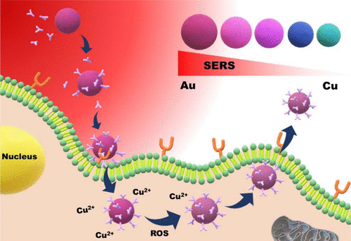
Here, we utilized inert Cu@poly(styrene-alt-maleic acid) (PSMA) nanoparticles (NPs) as a potential starting material, enabling restrictions on chemical etching and resistance to external toxic surfactant adsorption. Such modulated oxidation–dissolution of Cu allowed preservation of the original nano-Cu shape and facilitated the subsequent deposition of Au atoms to form AuCu nanoshells with a tunable Au/Cu ratio. An increase in the Au concentration fraction at the surface nanolayer of the AuxCu1–x nanoshells (x = 0.41–0.86) could intensify the polarizability at the interface structure to essentially aid both electromagnetic field- and chemical-improved surface-enhanced Raman scattering (SERS). Among the AuxCu1–x composites, Au0.86Cu0.14 nanoshells exhibited boost in SERS with amplification to 2–26-fold that of pure Au nanorods, Ag@ polyvinylpyrrolidone (PVP) NPs, Ag@PVP nanocubes, and AuyAg1–y@PSMA nanoshells. Because of the intended SERS response and lower cytotoxicity, we further conjugated the FGFR3 antibody onto the surface of the Au0.86Cu0.14 nanoshells to first demonstrate SERS detection for the highly selective sensing and recognition of T24 human bladder cells. Time-dependent SERS monitor presented the targeted labeling with a signal increase from 0 to 24 h followed by an endocytosis route. In bladder cancer cell uptake, the Au0.86Cu0.14 nanoshell possessed very slight release of Cu species which could deliver a “secretion signal” to trigger the outward transportation of AuCu hollow NPs to leave bladder living cells, showing an alternative method with the potential Au–Cu composite to overcome previous inorganic SERS NPs that often encounter cellular accumulation and cannot be secreted.
中文翻译:

空心Au x Cu 1– x合金纳米壳,用于基于表面的拉曼光谱跟踪膀胱癌细胞,然后触发可切除的分泌物
在这里,我们使用惰性的Cu @聚(苯乙烯ALT -马来酸)(PSMA)纳米颗粒(NP),其为潜在的起始原料,使上化学蚀刻和电阻限制到外部表面活性剂的毒性吸附。这种经过调节的铜的氧化溶解过程可以保留原始的纳米铜形状,并有助于随后的金原子沉积,形成具有可调金/铜比的金铜纳米壳。Au x Cu 1– x纳米壳的表面纳米层上的Au浓度分数的增加(x = 0.41-0.86)可能会增强界面结构的极化率,从根本上帮助电磁场和化学增强的表面增强拉曼散射(SERS)。在金XCu 1– x复合材料,Au 0.86 Cu 0.14纳米壳在SERS中表现出增强作用,增强了纯Au纳米棒,Ag @聚乙烯吡咯烷酮(PVP)NP,Ag @ PVP纳米立方体和Au y Ag 1– y @的2–26倍。PSMA纳米壳。由于预期的SERS反应和较低的细胞毒性,我们进一步将FGFR3抗体偶联到Au 0.86 Cu 0.14的表面上纳米壳首次展示了SERS检测对T24人膀胱细胞的高度选择性感应和识别。随时间变化的SERS监测器显示了靶向标记,信号从0到24小时增加,随后是胞吞途径。在膀胱癌细胞的摄取中,Au 0.86 Cu 0.14纳米壳具有非常轻微的Cu释放,可以释放“分泌信号”以触发AuCu中空NP的向外运输,从而离开膀胱活细胞,这显示了一种潜在的Au替代方法-Cu复合材料可克服以前的无机SERS NP,它们经常遇到细胞积累而无法分泌。
更新日期:2020-07-02
中文翻译:
空心Au x Cu 1– x合金纳米壳,用于基于表面的拉曼光谱跟踪膀胱癌细胞,然后触发可切除的分泌物
在这里,我们使用惰性的Cu @聚(苯乙烯ALT -马来酸)(PSMA)纳米颗粒(NP),其为潜在的起始原料,使上化学蚀刻和电阻限制到外部表面活性剂的毒性吸附。这种经过调节的铜的氧化溶解过程可以保留原始的纳米铜形状,并有助于随后的金原子沉积,形成具有可调金/铜比的金铜纳米壳。Au x Cu 1– x纳米壳的表面纳米层上的Au浓度分数的增加(x = 0.41-0.86)可能会增强界面结构的极化率,从根本上帮助电磁场和化学增强的表面增强拉曼散射(SERS)。在金XCu 1– x复合材料,Au 0.86 Cu 0.14纳米壳在SERS中表现出增强作用,增强了纯Au纳米棒,Ag @聚乙烯吡咯烷酮(PVP)NP,Ag @ PVP纳米立方体和Au y Ag 1– y @的2–26倍。PSMA纳米壳。由于预期的SERS反应和较低的细胞毒性,我们进一步将FGFR3抗体偶联到Au 0.86 Cu 0.14的表面上纳米壳首次展示了SERS检测对T24人膀胱细胞的高度选择性感应和识别。随时间变化的SERS监测器显示了靶向标记,信号从0到24小时增加,随后是胞吞途径。在膀胱癌细胞的摄取中,Au 0.86 Cu 0.14纳米壳具有非常轻微的Cu释放,可以释放“分泌信号”以触发AuCu中空NP的向外运输,从而离开膀胱活细胞,这显示了一种潜在的Au替代方法-Cu复合材料可克服以前的无机SERS NP,它们经常遇到细胞积累而无法分泌。


 京公网安备 11010802027423号