Computers in Biology and Medicine ( IF 7.7 ) Pub Date : 2020-07-01 , DOI: 10.1016/j.compbiomed.2020.103901 Danilo Samuel Jodas 1 , Maria Francisca Monteiro da Costa 2 , Tiago A A Parreira 3 , Aledir Silveira Pereira 4 , João Manuel R S Tavares 5
|
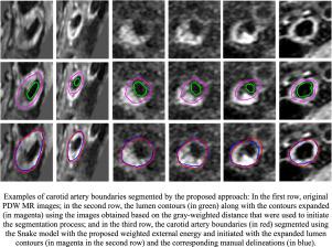
Segmentation methods have assumed an important role in image-based diagnosis of several cardiovascular diseases. Particularly, the segmentation of the boundary of the carotid artery is demanded in the detection and characterization of atherosclerosis and assessment of the disease progression. In this article, a fully automatic approach for the segmentation of the carotid artery boundary in Proton Density Weighted Magnetic Resonance Images is presented. The approach relies on the expansion of the lumen contour based on a distance map built using the gray-weighted distance relative to the center of the identified lumen region in the image under analysis. Then, a Snake model with a modified weighted external energy based on the combination of a balloon force along with a Gradient Vector Flow-based external energy is applied to the expanded contour towards the correct boundary of the carotid artery. The average values of the Dice coefficient, Polyline distance, mean contour distance and centroid distance found in the segmentation of 139 carotid arteries were 0.83 ± 0.11, 2.70 ± 1.69 pixels, 2.79 ± 1.89 pixels and 3.44 ± 2.82 pixels, respectively. The segmentation results of the proposed approach were also compared against the ones obtained by related approaches found in the literature, which confirmed the outstanding performance of the new approach. Additionally, the proposed weighted external energy for the Snake model was shown to be also robust to carotid arteries with large thickness and weak boundary image edges.
中文翻译:
在质子密度加权磁共振图像中,使用距离图和主动轮廓模型从管腔轮廓中分割出颈动脉边界。
分割方法在几种心血管疾病的基于图像的诊断中起着重要作用。特别地,在动脉粥样硬化的检测和表征以及疾病进展的评估中需要分割颈动脉的边界。在本文中,提出了一种在质子密度加权磁共振图像中分割颈动脉边界的全自动方法。该方法依赖于基于距离图的管腔轮廓扩展,该距离图是使用相对于所分析图像中已识别管腔区域中心的灰度加权距离构建的。然后,将基于气球力与基于梯度矢量流的外部能量相结合的,具有修改后的加权外部能量的Snake模型应用于朝向颈动脉正确边界的扩展轮廓。在139个颈动脉的分割中发现的Dice系数,折线距离,平均轮廓距离和质心距离的平均值分别为0.83±0.11、2.70±1.69像素,2.79±1.89像素和3.44±2.82像素。还将所提方法的分割结果与文献中相关方法获得的分割结果进行了比较,这证实了新方法的出色性能。另外,


 京公网安备 11010802027423号