Nanomedicine: Nanotechnology, Biology and Medicine ( IF 5.4 ) Pub Date : 2020-06-30 , DOI: 10.1016/j.nano.2020.102250 Sheng Zheng 1 , Ying Zhang 2 , Shujie Chen 2 , Zeyu Zhang 3 , Fei Chen 4 , Zizhen Zhang 4 , Zhenhua Hu 3 , Jie Tian 3 , Liangjing Wang 4
|
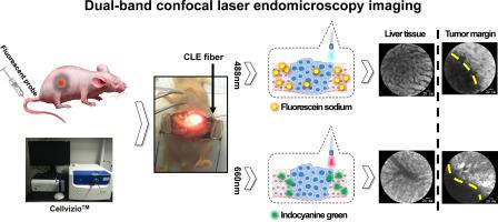
Accurate identification of tumor tissues and their margins are still challenging for conventional clinical imaging methods during liver cancer surgery. In this study, dual-band confocal laser endomicroscopy (CLE) combined with image mosaic was used to guide liver cancer surgery. In the experiments with mice bearing orthotropic liver tumor, CLE can accurately detect the tumors and identify their margins with two excitation wavelengths of 488 nm and 660 nm by clinically available dyes fluorescein sodium (FS) or indocyanine green (ICG). The mosaic CLE images enlarged the imaging field and detected the liver tumor margins more accurately. Normal liver tissues fluorescence intensity of CLE images was significantly higher than that of tumor tissues in the same tumor-bearing mice (P < 0.0001). Overall, dual-band CLE imaging demonstrates to be a promising method to identify liver tumor tissues and margins, which has the prospect of clinical application and helps to achieve intraoperative radical resection.
中文翻译:
双波段共聚焦激光内镜检查结合图像镶嵌技术在肝癌诊断中的初步研究。
对于肝癌手术期间的常规临床成像方法,准确识别肿瘤组织及其边缘仍然是挑战。在这项研究中,双波段共聚焦激光内镜检查(CLE)结合图像镶嵌技术被用于指导肝癌手术。在患有患有正交性肝肿瘤的小鼠的实验中,CLE可以通过临床上可用的荧光素钠(FS)或吲哚菁绿(ICG)准确地检测出肿瘤,并在488 nm和660 nm的两个激发波长下确定其边缘。马赛克CLE图像扩大了成像范围,并更准确地检测到肝肿瘤边缘。正常肝组织的CLE图像荧光强度显着高于同一只荷瘤小鼠的肿瘤组织(P <0.0001)。总体而言,双波段CLE成像被证明是鉴定肝脏肿瘤组织和切缘的一种有前途的方法,具有临床应用前景并有助于实现术中根治性切除。


 京公网安备 11010802027423号