Our official English website, www.x-mol.net, welcomes your feedback! (Note: you will need to create a separate account there.)
Schwann cells selectively myelinate primary motor axons via neuregulin-ErbB signaling.
Glia ( IF 6.2 ) Pub Date : 2020-06-26 , DOI: 10.1002/glia.23871 Dong-Won Lee 1 , Eunmi Kim 1 , Inyoung Jeong 1 , Hwan-Ki Kim 1 , Suhyun Kim 1 , Hae-Chul Park 1
Glia ( IF 6.2 ) Pub Date : 2020-06-26 , DOI: 10.1002/glia.23871 Dong-Won Lee 1 , Eunmi Kim 1 , Inyoung Jeong 1 , Hwan-Ki Kim 1 , Suhyun Kim 1 , Hae-Chul Park 1
Affiliation
|
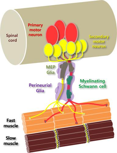
Spinal motor neurons project their axons out of the spinal cord via the motor exit point (MEP) and regulate their target muscle fibers for diverse behaviors. Several populations of glial cells including Schwann cells, MEP glia, and perineurial glia are tightly associated with spinal motor axons in nerve fascicles. Zebrafish have two types of spinal motor neurons, primary motor neurons (PMNs) and secondary motor neurons (SMNs). PMNs are implicated in the rapid response, whereas SMNs are implicated in normal and slow movements. However, the precise mechanisms mediating the distinct functions of PMNs and SMNs in zebrafish are unclear. In this study, we found that PMNs were myelinated by MEP glia and Schwann cells, whereas SMNs remained unmyelinated at the examined stages. Immunohistochemical analysis revealed that myelinated PMNs solely innervated fast muscle through a distributed neuromuscular junction (NMJ), whereas unmyelinated SMNs innervated both fast and slow muscle through distributed and myoseptal NMJs, respectively, indicating that myelinated PMNs could provide rapid responses for startle and escape movements, while unmyelinated SMNs regulated normal, slow movement. Further, we demonstrate that neuregulin 1 (Nrg1) type III‐ErbB signaling provides a key instructive signal that determines the myelination of primary motor axons by MEP glia and Schwann cells. Perineurial glia ensheathed unmyelinated secondary motor axons and myelinated primary motor nerves. Ensheathment required interaction with both MEP glia and Schwann cells. Collectively, these data suggest that primary and secondary motor neurons contribute to the regulation of movement in zebrafish with distinct patterns of myelination.
中文翻译:

雪旺氏细胞通过神经调节蛋白-ErbB 信号选择性地髓鞘化初级运动轴突。
脊髓运动神经元通过运动出口点 (MEP) 将其轴突投射到脊髓外,并调节其目标肌肉纤维以实现不同的行为。包括雪旺氏细胞、MEP 神经胶质和神经周围神经胶质在内的几种神经胶质细胞群与神经束中的脊髓运动轴突紧密相关。斑马鱼有两种类型的脊髓运动神经元,初级运动神经元 (PMN) 和次级运动神经元 (SMN)。PMN 与快速反应有关,而 SMN 与正常和缓慢运动有关。然而,在斑马鱼中介导 PMN 和 SMN 不同功能的精确机制尚不清楚。在这项研究中,我们发现中性粒细胞由 MEP 神经胶质细胞和雪旺细胞有髓鞘,而 SMN 在检查阶段保持无髓鞘。免疫组织化学分析显示,有髓鞘的中性粒细胞仅通过分布式神经肌肉接头(NMJ)支配快肌,而无髓鞘的中性粒细胞分别通过分布式神经肌肉接头和肌间隔NMJ支配快肌和慢肌,表明有髓中性粒细胞可以为惊吓和逃逸运动提供快速反应,而无髓鞘的 SMN 调节正常、缓慢的运动。此外,我们证明了神经调节蛋白 1 (Nrg1) 型 III-ErbB 信号提供了一个关键的指导性信号,该信号决定了 MEP 神经胶质细胞和雪旺氏细胞对初级运动轴突的髓鞘形成。神经周围神经胶质包裹着无髓鞘的次级运动轴突和有髓鞘的初级运动神经。Ensheathment 需要与 MEP 神经胶质细胞和雪旺氏细胞相互作用。总的来说,
更新日期:2020-06-26
中文翻译:
雪旺氏细胞通过神经调节蛋白-ErbB 信号选择性地髓鞘化初级运动轴突。
脊髓运动神经元通过运动出口点 (MEP) 将其轴突投射到脊髓外,并调节其目标肌肉纤维以实现不同的行为。包括雪旺氏细胞、MEP 神经胶质和神经周围神经胶质在内的几种神经胶质细胞群与神经束中的脊髓运动轴突紧密相关。斑马鱼有两种类型的脊髓运动神经元,初级运动神经元 (PMN) 和次级运动神经元 (SMN)。PMN 与快速反应有关,而 SMN 与正常和缓慢运动有关。然而,在斑马鱼中介导 PMN 和 SMN 不同功能的精确机制尚不清楚。在这项研究中,我们发现中性粒细胞由 MEP 神经胶质细胞和雪旺细胞有髓鞘,而 SMN 在检查阶段保持无髓鞘。免疫组织化学分析显示,有髓鞘的中性粒细胞仅通过分布式神经肌肉接头(NMJ)支配快肌,而无髓鞘的中性粒细胞分别通过分布式神经肌肉接头和肌间隔NMJ支配快肌和慢肌,表明有髓中性粒细胞可以为惊吓和逃逸运动提供快速反应,而无髓鞘的 SMN 调节正常、缓慢的运动。此外,我们证明了神经调节蛋白 1 (Nrg1) 型 III-ErbB 信号提供了一个关键的指导性信号,该信号决定了 MEP 神经胶质细胞和雪旺氏细胞对初级运动轴突的髓鞘形成。神经周围神经胶质包裹着无髓鞘的次级运动轴突和有髓鞘的初级运动神经。Ensheathment 需要与 MEP 神经胶质细胞和雪旺氏细胞相互作用。总的来说,


 京公网安备 11010802027423号