Journal of the Mechanical Behavior of Biomedical Materials ( IF 3.9 ) Pub Date : 2020-06-25 , DOI: 10.1016/j.jmbbm.2020.103887 Yoshihiro Obata 1 , Hrishikesh A Bale 2 , Harold S Barnard 3 , Dula Y Parkinson 3 , Tamara Alliston 4 , Claire Acevedo 5
|
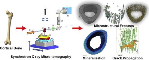
All levels of the unique hierarchical structure of bone, consisting of collagen and hydroxyapatite crystals at the nanoscale to osteon/lamellae structures at the microscale, contribute to its characteristic toughness and material properties. Elements of bone's density and size contribute to bone quantity (or bone mass), whereas elements of bone's material composition, material properties, internal structure, and organization describe bone quality. Bone quantity and quality can be degraded by factors such as aging, disease, treatments, and irradiation, compromising its ability to resist fracture and sustain loading. Accessing the morphology and architecture of bone at the microscale to quantify microstructural features and assess the degree of mineralization and path of crack propagation in bone provides crucial information on how these factors are influencing bone quantity and quality. Synchrotron radiation micro-computed tomography (SRμCT) was first used to assess bone structure at the end of the 1990's. One of the main advantages of the technique is that it enables accurate three-dimensional (3D), non-destructive quantification of structure while traditional histomorphometry on histological sections is inherantly destructive to the sample and two-dimensional (2D). Additionally, SRμCT uses monochromatic, high-flux X-ray beams to provide high-resolution and high-contrast imaging of bone samples. This allows the quantification of small microstructural features (e.g. osteocyte lacunae, canals, trabeculae, microcracks) and direct gray value compositional mapping (e.g. mineral quantification, cement lines) with greater speed and fidelity than lab-based micro-computed tomography. In this article, we review how SRμCT has been applied to bone research to elucidate the mechanisms by which bone aging, disease, and other factors affect bone fragility and resistance to fracture.
中文翻译:
定量和定性的骨成像:骨研究中同步辐射显微断层分析的综述。
骨骼的独特层次结构的所有级别(从纳米级的胶原蛋白和羟基磷灰石晶体到微米级的骨/片状结构)都有助于其特性韧性和材料性能。骨骼的密度和大小元素影响骨骼的数量(或骨骼质量),而骨骼的材料组成,材料特性,内部结构和组织元素描述骨骼质量。骨骼的数量和质量会因诸如衰老,疾病,治疗和照射等因素而降低,从而损害其抵抗骨折和承受负荷的能力。在微观尺度上访问骨骼的形态和结构以量化微观结构特征并评估矿化程度和裂缝在骨骼中的传播路径,提供了有关这些因素如何影响骨骼数量和质量的重要信息。在1990年代末,同步辐射微型计算机断层扫描(SRμCT)首次用于评估骨骼结构。该技术的主要优势之一是,它可以对结构进行精确的三维(3D)无损量化,而组织学切片上的传统组织形态学对样本和二维(2D)则具有固有的破坏性。此外,SRμCT使用单色的高通量X射线束来提供骨骼样品的高分辨率和高对比度成像。与基于实验室的微型计算机断层扫描相比,这可以对较小的微结构特征(例如骨细胞腔,运河,小梁,微裂纹)进行定量,并可以直接灰度值映射(例如矿物质定量,水泥线),且速度和保真度更高。在本文中,我们回顾了SRμCT如何应用于骨骼研究,以阐明骨骼老化,疾病和其他因素影响骨骼脆弱性和抗断裂性的机制。


 京公网安备 11010802027423号