当前位置:
X-MOL 学术
›
ACS Chem. Neurosci.
›
论文详情
Our official English website, www.x-mol.net, welcomes your feedback! (Note: you will need to create a separate account there.)
Brain Endothelial Cell-Derived Exosomes Induce Neuroplasticity in Rats with Ischemia/Reperfusion Injury.
ACS Chemical Neuroscience ( IF 5 ) Pub Date : 2020-06-23 , DOI: 10.1021/acschemneuro.0c00089 Beiyao Gao 1 , Shaoting Zhou 2 , Chengcheng Sun 3 , Dandan Cheng 1 , Ye Zhang 3 , Xutong Li 2 , Li Zhang 1 , Jing Zhao 2 , Dongsheng Xu 4, 5, 6 , Yulong Bai 1
ACS Chemical Neuroscience ( IF 5 ) Pub Date : 2020-06-23 , DOI: 10.1021/acschemneuro.0c00089 Beiyao Gao 1 , Shaoting Zhou 2 , Chengcheng Sun 3 , Dandan Cheng 1 , Ye Zhang 3 , Xutong Li 2 , Li Zhang 1 , Jing Zhao 2 , Dongsheng Xu 4, 5, 6 , Yulong Bai 1
Affiliation
|
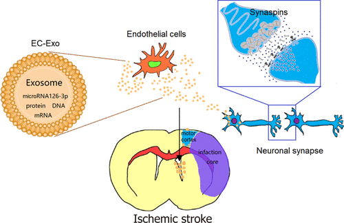
Exosomes derived from the cerebral endothelial cells play essential roles in protecting neurons from hypoxia injury, but little is known regarding the biological effects and mechanisms of exosomes on brain plasticity. In this study, exosomes were isolated from rodent cerebral endothelial cells (bEnd.3 cells) by ultracentrifugation, either endothelial cell-derived exosomes (EC-Exo) or PBS was injected intraventricularly 2 h after the middle cerebral artery occlusion/reperfusion (MCAO/R) model surgery in the Exo group and control group, respectively. Sham group rats received the same surgical but not ischemic procedure. We evaluated the motor function of rats after MCAO/R, and the foot-fault rate of the Exo group was significantly lower than that of the control group within 23 days (p < 0.05); the Catwalk analysis also showed gait difference between two groups (p < 0.05). On day 28 after MCAO/R, we euthanized the rats, removed the motor cortex from the brain, and then sequenced the genes by using GO and KEGG to find transcriptome analysis of biological terms and functional annotations: The pathway enrichment revealed that the function of synaptic transmission, regulation of synaptic plasticity, and regulation of synaptic vesicle cycle was significantly enriched with the Exo group than control group. Furthermore, the upregulation of synapsin-I expression in the motor cortex (p < 0.05) as well as the increase of the length of the dendrites were found in the Exo group (p < 0.05) than the control group. We determined the content of exosome microRNA levels, and microRNA-126-3p was the highest (TPM) by transcriptome analysis. Moreover, the microRNA-126-3p protected PC12 cells from apoptosis and increased neurite outgrowth, illustrating the mechanism of how exosomes play a role in altering brain plasticity. This study demonstrated that EC-Exo promoted functional motor recovery in the MCAO/R model, exosomes were critical for the reconstruction of synaptic function in ischemic brain injury, and microRNA-126-3p from EC-Exo could serve as a treatment for nerve damage.
中文翻译:

脑内皮细胞衍生的外来体在缺血/再灌注损伤大鼠中诱导神经可塑性。
衍生自脑内皮细胞的外泌体在保护神经元免受缺氧损伤中起着重要作用,但关于外泌体对大脑可塑性的生物学作用和机制知之甚少。在这项研究中,通过超速离心从啮齿动物的大脑内皮细胞(bEnd.3细胞)中分离出外泌体,在大脑中动脉闭塞/再灌注(MCAO /)后2小时内,从内皮细胞衍生的外泌体(EC-Exo)或PBS注射R)分别在Exo组和对照组中进行模型手术。假手术组大鼠接受相同的外科手术但不接受缺血程序。我们评估了MCAO / R后大鼠的运动功能,Exo组的足部断脚率在23天内显着低于对照组(p<0.05); Catwalk分析还显示了两组之间的步态差异(p <0.05)。MCAO / R后第28天,我们对大鼠实施安乐死,从大脑中取出运动皮层,然后使用GO和KEGG对基因进行测序,以寻找生物学术语和功能注释的转录组分析:途径富集表明, Exo组比对照组显着丰富了突触传递,突触可塑性调节和突触小泡周期调节。此外,在Exo组中发现运动皮层中突触素-I表达的上调(p <0.05)以及树突长度的增加(p<0.05)。我们确定了外泌体microRNA水平的含量,通过转录组分析,microRNA-126-3p最高(TPM)。此外,microRNA-126-3p保护PC12细胞免于凋亡和神经突增生,说明了外泌体如何在改变大脑可塑性中发挥作用的机制。这项研究表明EC-Exo促进了MCAO / R模型中的功能性运动恢复,外来体对于缺血性脑损伤突触功能的重建至关重要,而EC-Exo的microRNA-126-3p可以作为神经损伤的治疗方法。
更新日期:2020-08-05
中文翻译:
脑内皮细胞衍生的外来体在缺血/再灌注损伤大鼠中诱导神经可塑性。
衍生自脑内皮细胞的外泌体在保护神经元免受缺氧损伤中起着重要作用,但关于外泌体对大脑可塑性的生物学作用和机制知之甚少。在这项研究中,通过超速离心从啮齿动物的大脑内皮细胞(bEnd.3细胞)中分离出外泌体,在大脑中动脉闭塞/再灌注(MCAO /)后2小时内,从内皮细胞衍生的外泌体(EC-Exo)或PBS注射R)分别在Exo组和对照组中进行模型手术。假手术组大鼠接受相同的外科手术但不接受缺血程序。我们评估了MCAO / R后大鼠的运动功能,Exo组的足部断脚率在23天内显着低于对照组(p<0.05); Catwalk分析还显示了两组之间的步态差异(p <0.05)。MCAO / R后第28天,我们对大鼠实施安乐死,从大脑中取出运动皮层,然后使用GO和KEGG对基因进行测序,以寻找生物学术语和功能注释的转录组分析:途径富集表明, Exo组比对照组显着丰富了突触传递,突触可塑性调节和突触小泡周期调节。此外,在Exo组中发现运动皮层中突触素-I表达的上调(p <0.05)以及树突长度的增加(p<0.05)。我们确定了外泌体microRNA水平的含量,通过转录组分析,microRNA-126-3p最高(TPM)。此外,microRNA-126-3p保护PC12细胞免于凋亡和神经突增生,说明了外泌体如何在改变大脑可塑性中发挥作用的机制。这项研究表明EC-Exo促进了MCAO / R模型中的功能性运动恢复,外来体对于缺血性脑损伤突触功能的重建至关重要,而EC-Exo的microRNA-126-3p可以作为神经损伤的治疗方法。


 京公网安备 11010802027423号