Journal of Neuroscience Methods ( IF 3 ) Pub Date : 2020-06-21 , DOI: 10.1016/j.jneumeth.2020.108825 Matthias Weigel 1 , Tanja Haas 2 , Maria Janina Wendebourg 3 , Regina Schlaeger 3 , Oliver Bieri 4
|
Background
Spinal cord (SC) gray and white matter atrophy quantification by advanced morphometric MRI can help to better characterize the course of neurodegenerative diseases in vivo, such as e.g. lower motor neuron disorders. Imaging the lower thoracic cord - containing those motor neurons that control leg function - could be particularly informative, however, is challenging due to tissue composition, physiological motion and large field of views.
New method
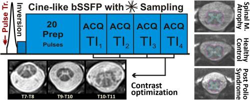
An “averaged magnetization inversion recovery acquisitions” (AMIRA) approach with a radial k-space acquisition scheme was developed. The method is designed for morphometric SC imaging with a focus on the thoracic SC.
Results
In a typical setting, radial AMIRA acquires transverse slices with a high 0.50 × 0.50mm2 in-plane resolution and a pronounced positive contrast between thoracic gray and white matter, within typically 2:39 min. Additional proof-of-concept measurements in patients demonstrate that such contrast and resolving capability is indeed necessary to assess potential atrophy of the anterior horns.
Comparison with existing method(s)
Radial AMIRA utilizes two benefits of radial MRI techniques: being generally less prone to motion effects and that fold over artifacts can manifest less intrusively. These benefits are united with the original AMIRA approach which allows the contrast to be ‘tuned’ and improved based on the combination of five simultaneously acquired images of different tissue contrast.
Conclusions
Radial AMIRA is a promising approach for in vivo SC gray and white matter atrophy visualization and quantification in lower motor neuron diseases and other autoimmune or genetic diseases involving the entire (not only cervical) spinal cord.
中文翻译:
使用放射状采样的平均磁化反转恢复采集对胸脊髓成像。
背景
通过先进的形态学MRI量化脊髓(SC)的灰质和白质萎缩可以帮助更好地表征体内神经退行性疾病的病程,例如下运动神经元疾病。包含控制腿部功能的运动神经元的下胸索成像可能特别有用,但是由于组织组成,生理运动和大视野,这具有挑战性。
新方法
开发了一种具有径向k空间采集方案的“平均磁化反演恢复采集”(AMIRA)方法。该方法专为形态SC成像而设计,重点是胸SC。
结果
在典型情况下,放射状AMIRA通常在2:39分钟内即可获得0.50×0.50mm 2的高平面分辨率和胸灰与白质之间明显正对比度的横向切片。在患者中进行的其他概念验证测量表明,这种对比度和分辨能力确实是评估潜在的前角萎缩所必需的。
与现有方法的比较
放射状AMIRA利用放射状MRI技术的两个好处:通常不易受到运动影响,并且折叠在伪影上的侵入性也较小。这些好处与原始AMIRA方法结合在一起,该方法允许基于五个同时获取的不同组织对比度的图像的组合来“调整”和改善对比度。
结论
放射状AMIRA是一种有希望的方法,可用于在下运动神经元疾病以及涉及整个(不仅是颈椎)脊髓的其他自身免疫或遗传疾病中进行体内SC灰质和白质萎缩的可视化和定量。


 京公网安备 11010802027423号