当前位置:
X-MOL 学术
›
Biomacromolecules
›
论文详情
Our official English website, www.x-mol.net, welcomes your feedback! (Note: you will need to create a separate account there.)
Plasmonic Enhancement of Two-Photon Excitation Fluorescence by Colloidal Assemblies of Very Small AuNPs Templated on M13 Phage.
Biomacromolecules ( IF 6.2 ) Pub Date : 2020-06-18 , DOI: 10.1021/acs.biomac.0c00401 Esen Sokullu 1 , Maxime Pinsard 1 , Jiawei Zhang 1 , Julien Plathier 1 , Gitanjali Kolhatkar 1 , Amy Szuchmacher Blum 2 , François Légaré 1 , Andreas Ruediger 1 , Tsuneyuki Ozaki 1 , Marc A Gauthier 1
Biomacromolecules ( IF 6.2 ) Pub Date : 2020-06-18 , DOI: 10.1021/acs.biomac.0c00401 Esen Sokullu 1 , Maxime Pinsard 1 , Jiawei Zhang 1 , Julien Plathier 1 , Gitanjali Kolhatkar 1 , Amy Szuchmacher Blum 2 , François Légaré 1 , Andreas Ruediger 1 , Tsuneyuki Ozaki 1 , Marc A Gauthier 1
Affiliation
|
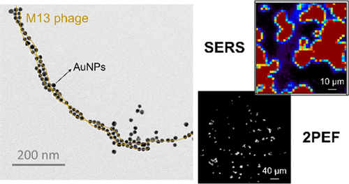
In this study, an engineered M13 bacteriophage was examined as a biological template to create a well-defined spacing between very small gold nanoparticles (AuNPs 3–13 nm). The effect of the AuNP particle size on the enhancement of the nonlinear process of two-photon excitation fluorescence (2PEF) was investigated. Compared to conventional (one-photon) microscopy techniques, such nonlinear processes are less susceptible to scattering given that the density of background-scattered photons is too low to generate a detectable signal. Besides this, the use of very small AuNPs in 2PEF microscopy becomes more advantageous because individual “isolated” AuNPs of this size do not sufficiently enhance 2PEF to produce a detectable signal, resulting in even less background signal. To investigate the 2PEF of the AuNP–M13 assemblies, a variety of sample preparation approaches are tested, and surface-enhanced Raman spectroscopy (SERS) is employed to study the strength of plasmon coupling within the gaps of AuNPs assembled on the M13 template. Results indicate that assemblies prepared with 9–13 nm AuNP were able to clearly label Escherichia coli cells and produce a 2PEF signal that was orders of magnitude higher than the isolated AuNP (below the threshold of detection). This study thus provides a better understanding of the opportunities and limitations relevant to the use of such small AuNPs within colloidal plasmonic assemblies, for applications in biodetection or as imaging contrast agents.
中文翻译:

通过在M13噬菌体上模板化的非常小的AuNP的胶体组装对两光子激发荧光的等离子体增强。
在这项研究中,将工程化的M13噬菌体作为一种生物模板进行了检查,以在非常小的金纳米颗粒(AuNPs 3–13 nm)之间建立明确的间隔。研究了AuNP粒径对增强双光子激发荧光(2PEF)非线性过程的影响。与传统的(单光子)显微镜技术相比,由于背景散射光子的密度太低而无法生成可检测的信号,因此此类非线性过程不易受到散射的影响。除此之外,在2PEF显微镜中使用非常小的AuNP变得更加有利,因为这种大小的单个“分离的” AuNP无法充分增强2PEF以产生可检测的信号,从而导致更少的背景信号。要研究AuNP–M13组件的2PEF,测试了多种样品制备方法,并使用表面增强拉曼光谱(SERS)研究了在M13模板上组装的AuNP间隙内的等离激元耦合强度。结果表明,使用9-13 nm AuNP制备的组件能够清楚地标记大肠杆菌细胞并产生2PEF信号,该信号比分离的AuNP高几个数量级(低于检测阈值)。因此,这项研究可以更好地理解与在胶体等离子体组件中使用这种小的AuNPs有关的机会和局限性,以用于生物检测或成像造影剂。
更新日期:2020-07-13
中文翻译:
通过在M13噬菌体上模板化的非常小的AuNP的胶体组装对两光子激发荧光的等离子体增强。
在这项研究中,将工程化的M13噬菌体作为一种生物模板进行了检查,以在非常小的金纳米颗粒(AuNPs 3–13 nm)之间建立明确的间隔。研究了AuNP粒径对增强双光子激发荧光(2PEF)非线性过程的影响。与传统的(单光子)显微镜技术相比,由于背景散射光子的密度太低而无法生成可检测的信号,因此此类非线性过程不易受到散射的影响。除此之外,在2PEF显微镜中使用非常小的AuNP变得更加有利,因为这种大小的单个“分离的” AuNP无法充分增强2PEF以产生可检测的信号,从而导致更少的背景信号。要研究AuNP–M13组件的2PEF,测试了多种样品制备方法,并使用表面增强拉曼光谱(SERS)研究了在M13模板上组装的AuNP间隙内的等离激元耦合强度。结果表明,使用9-13 nm AuNP制备的组件能够清楚地标记大肠杆菌细胞并产生2PEF信号,该信号比分离的AuNP高几个数量级(低于检测阈值)。因此,这项研究可以更好地理解与在胶体等离子体组件中使用这种小的AuNPs有关的机会和局限性,以用于生物检测或成像造影剂。


 京公网安备 11010802027423号