当前位置:
X-MOL 学术
›
ACS Chem. Biol.
›
论文详情
Our official English website, www.x-mol.net, welcomes your feedback! (Note: you will need to create a separate account there.)
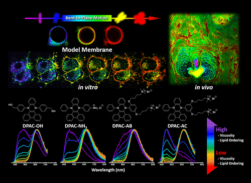
Fluorescence Probes Exhibit Photoinduced Structural Planarization: Sensing In Vitro and In Vivo Microscopic Dynamics of Viscosity Free from Polarity Interference.
ACS Chemical Biology ( IF 4 ) Pub Date : 2020-06-16 , DOI: 10.1021/acschembio.0c00100 Cheng-Ham Wu,Yi Chen,Kyrylo A Pyrshev,Yi-Ting Chen,Zhiyun Zhang,Kai-Hsin Chang,Semen O Yesylevskyy,Alexander P Demchenko,Pi-Tai Chou
ACS Chemical Biology ( IF 4 ) Pub Date : 2020-06-16 , DOI: 10.1021/acschembio.0c00100 Cheng-Ham Wu,Yi Chen,Kyrylo A Pyrshev,Yi-Ting Chen,Zhiyun Zhang,Kai-Hsin Chang,Semen O Yesylevskyy,Alexander P Demchenko,Pi-Tai Chou
|
We demonstrate the construction of wavelength λ-ratiometric images that allow visualizing the distribution of microscopic dynamics within living cells and tissues by using the newly developed principle of fluorescence response. The bent-to-planar motion in the excited state of incorporated fluorescence probes leads to elongation of the π-delocalization, resulting in microviscosity-dependent but polarity-insensitive interplay between well-separated blue and red bands in emission spectra. This allows constructing the exceptionally contrasted images of cellular dynamics. Moreover, the application of probes with increased affinity toward biological membranes allowed detecting the differences in dynamics between the plasma membrane and intracellular membrane structures. Such λ-ratiometric microviscosity imaging was extended for mapping the living tissues and observing their inflammation-dependent changes.
中文翻译:

荧光探针展示出光诱导的结构平面化:体外和体内的微观粘度动态传感,不受极性干扰。
我们演示了波长λ比例图像的构建,该图像允许通过使用新开发的荧光反应原理可视化活细胞和组织内的微观动力学分布。掺入的荧光探针在激发态下的平面弯曲运动导致π离域的延长,从而导致发射光谱中分离良好的蓝带和红带之间具有微粘度依赖性,但对极性不敏感。这允许构造细胞动力学的异常对比的图像。而且,对生物膜具有增加的亲和力的探针的应用允许检测质膜和细胞内膜结构之间的动力学差异。
更新日期:2020-07-17
中文翻译:
荧光探针展示出光诱导的结构平面化:体外和体内的微观粘度动态传感,不受极性干扰。
我们演示了波长λ比例图像的构建,该图像允许通过使用新开发的荧光反应原理可视化活细胞和组织内的微观动力学分布。掺入的荧光探针在激发态下的平面弯曲运动导致π离域的延长,从而导致发射光谱中分离良好的蓝带和红带之间具有微粘度依赖性,但对极性不敏感。这允许构造细胞动力学的异常对比的图像。而且,对生物膜具有增加的亲和力的探针的应用允许检测质膜和细胞内膜结构之间的动力学差异。


 京公网安备 11010802027423号