Molecular Therapy - Methods & Clinical Development ( IF 4.7 ) Pub Date : 2020-06-15 , DOI: 10.1016/j.omtm.2020.06.008 Johannes Winkler 1 , Dominika Lukovic 1 , Julia Mester-Tonczar 1 , Katrin Zlabinger 1 , Alfred Gugerell 1 , Noemi Pavo 1 , András Jakab 2, 3 , Zsuzsanna Szankai 2 , Denise Traxler 1 , Claudia Müller 1 , Andreas Spannbauer 1 , Martin Riesenhuber 1 , Ena Hašimbegović 1 , James Dawkins 4 , Matthias Zimmermann 5 , Hendrik J Ankersmit 5 , Eduardo Marbán 4 , Mariann Gyöngyösi 1
|
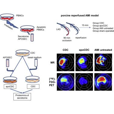
Cardiosphere-derived cells (CDCs) are progenitor cells derived from heart tissue and have shown promising results in preclinical models. APOSEC, the secretome of irradiated peripheral blood mononuclear cells, has decreased infarct size in acute and chronic experimental myocardial infarction (MI). We enhanced the effect of CDCs with APOSEC preconditioning (apoCDC) and investigated the reparative effect in a translational pig model of reperfused MI. Supernatants of CDCs, assessed by proteomic analysis, revealed reduced production of extracellular matrix proteins after in vitro APOSEC preconditioning. In a porcine model of catheter-based reperfused anterior acute MI (AMI), CDCs with (apoCDC, n = 8) or without APOSEC preconditioning (CDC, n = 6) were infused intracoronary, 15 min after the start of reperfusion. Untreated AMI animals (n = 7) and sham procedures (n = 5) functioned as controls. 2-deoxy-2-(18 F)-fluoro-D-glucose-positron emission tomography-magnetic resonance imaging ([18F]FDG-PET-MRI), with late enhancement after 1 month, showed reduced scar volume and lower transmurality of the infarcted area in CDC and apoCDC compared to AMI controls. Segmental quantitative PET images displayed indicated more residual viability in apoCDC. The left-ventricle (LV) ejection fraction was improved nonsignificantly to 45.8% ± 8.6% for apoCDC and 43.5% ± 7.1% for CDCs compared to 38.5% ± 4.4% for untreated AMI. Quantitative hybrid [18F]FDG-PET-MRI demonstrated improved metabolic and functional recovery after CDC administration, whereas apoCDCs induced preservation of viability of the infarcted area.
中文翻译:
定量混合心脏[18F] FDG-PET-MRI图像,用于评估预处理的心球衍生细胞的心脏修复。
心球来源的细胞(CDC)是源自心脏组织的祖细胞,并且在临床前模型中显示出令人鼓舞的结果。在急性和慢性实验性心肌梗塞(MI)中,受辐照的外周血单核细胞的分泌组APOSEC的梗塞面积已减小。我们使用APOSEC预处理(apoCDC)增强了CDC的作用,并研究了在再灌注MI的翻译猪模型中的修复作用。通过蛋白质组学分析评估的CDC上清液显示体外培养后细胞外基质蛋白的产生减少APOSEC预处理。在以猪为基础的再灌注前急性急性心肌梗死(AMI)的猪模型中,在再灌注开始后15分钟,将具有(apoCDC,n = 8)或不具有APOSEC预处理(CDC,n = 6)的CDC注入冠状动脉内。未经治疗的AMI动物(n = 7)和假手术(n = 5)作为对照。2-脱氧-2-(18 F)-氟-D-葡萄糖-正电子发射断层扫描-磁共振成像([ 18F] FDG-PET-MRI)与AMI对照相比,CDC和apoCDC的梗塞区域疤痕量减少,梗死区域的透壁性降低,但在1个月后才有所增强。显示的分段定量PET图像表明apoCDC中有更多的残留活力。与apoCDC和CDC相比,左心室(LV)射血分数无明显改善,分别为45.8%±8.6%和43.5%±7.1%,而未经治疗的AMI为38.5%±4.4%。定量杂交[ 18 F] FDG-PET-MRI证明,施用CDC后代谢和功能恢复得到改善,而apoCDC诱导了梗塞区域活力的保留。


 京公网安备 11010802027423号