当前位置:
X-MOL 学术
›
Environ. Sci.: Nano
›
论文详情
Our official English website, www.x-mol.net, welcomes your feedback! (Note: you will need to create a separate account there.)
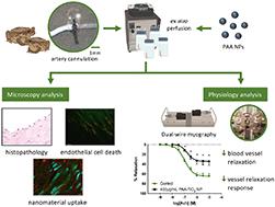
Polyacrylic acid coated nanoparticles elicit endothelial cell apoptosis and diminish vascular relaxation in ex vivo perfused iliac arteries of the cane toad (Rhinella marina)
Environmental Science: Nano ( IF 7.3 ) Pub Date : 2020-06-12 , DOI: 10.1039/d0en00229a Van A. Ortega 1, 2, 3, 4, 5 , Melissa S. Cameron 6, 7, 8, 9 , James L. Stafford 1, 2, 3, 4 , Greg G. Goss 1, 2, 3, 4, 10 , John A. Donald 9, 11, 12, 13 , Aaron G. Schultz 9, 11, 12, 13
Environmental Science: Nano ( IF 7.3 ) Pub Date : 2020-06-12 , DOI: 10.1039/d0en00229a Van A. Ortega 1, 2, 3, 4, 5 , Melissa S. Cameron 6, 7, 8, 9 , James L. Stafford 1, 2, 3, 4 , Greg G. Goss 1, 2, 3, 4, 10 , John A. Donald 9, 11, 12, 13 , Aaron G. Schultz 9, 11, 12, 13
Affiliation
|
Nanoparticles (NP) that penetrate external protective barriers of an animal are transported and distributed to various tissues and organs via the circulatory system. Whilst much attention has been dedicated to NP effects on target organs, surprisingly few studies have investigated effects on the vascular system itself, despite the critical role it plays in transporting red and white blood cells, nutrients, as well as coordinating many physiological responses, like immunity and stress. In this study we perfused iliac arteries extracted from cane toads (Rhinella marina) with polyacrylic acid (PAA) coated TiO2 NPs (3–9 nm) to directly assess effects on uptake across luminal endothelial layers, changes to vessel physiological function and toxicity. Perfusion of the iliac artery with 400 μg mL−1 PAA-TiO2 NPs resulted in histopathological changes in the vascular endothelial layer due to increased endothelial cell death via apoptosis. The PAA-NPs were observed to penetrate at least two cell layers and were present in both endothelial cells and underlying smooth muscle cells. Dual-wire myography experiments revealed that the highest dose of PAA-TiO2 NPs significantly inhibited the ACh-mediated vasodilation in blood vessels by ∼50%, which appeared to be attributable to the damaged endothelial layer in the lumen of the vessels. This is the first study to use an ex vivo perfusion method that mimics the blood circulation coupled with dual-wire myography to demonstrate the adverse effects of NPs on blood vessel physiology.
中文翻译:

涂有聚丙烯酸的纳米颗粒在蟾蜍蟾蜍离体灌注的动脉中引起内皮细胞凋亡并减少血管舒张。
穿透动物外部保护屏障的纳米颗粒(NP)通过循环系统运输并分布到各种组织和器官。尽管人们一直将注意力集中在NP对靶器官的影响上,但令人惊讶的是,很少有研究研究对NP的影响,尽管它在运输红细胞和白细胞,营养以及协调许多生理反应方面起着关键作用,例如免疫力和压力。在这项研究中,我们用涂有聚丙烯酸(PAA)的TiO 2灌注了从甘蔗蟾蜍(Rhinella marina)提取的ilia动脉。NP(3–9 nm)可直接评估对腔内内皮层摄取,血管生理功能和毒性变化的影响。用400微克毫升髂动脉灌注-1 PAA-的TiO 2纳米粒子导致了血管内皮层由于增加的内皮细胞死亡的组织病理学改变经由凋亡。观察到PAA-NP穿透至少两个细胞层并且存在于内皮细胞和下面的平滑肌细胞中。双线肌成像实验显示最高剂量的PAA-TiO 2NPs显着抑制血管中ACh介导的血管舒张约50%,这似乎归因于血管腔中内皮层的破坏。这是第一个使用体外灌注方法模拟血液循环的研究,结合双线肌成像技术来证明NP对血管生理的不利影响。
更新日期:2020-07-16
中文翻译:
涂有聚丙烯酸的纳米颗粒在蟾蜍蟾蜍离体灌注的动脉中引起内皮细胞凋亡并减少血管舒张。
穿透动物外部保护屏障的纳米颗粒(NP)通过循环系统运输并分布到各种组织和器官。尽管人们一直将注意力集中在NP对靶器官的影响上,但令人惊讶的是,很少有研究研究对NP的影响,尽管它在运输红细胞和白细胞,营养以及协调许多生理反应方面起着关键作用,例如免疫力和压力。在这项研究中,我们用涂有聚丙烯酸(PAA)的TiO 2灌注了从甘蔗蟾蜍(Rhinella marina)提取的ilia动脉。NP(3–9 nm)可直接评估对腔内内皮层摄取,血管生理功能和毒性变化的影响。用400微克毫升髂动脉灌注-1 PAA-的TiO 2纳米粒子导致了血管内皮层由于增加的内皮细胞死亡的组织病理学改变经由凋亡。观察到PAA-NP穿透至少两个细胞层并且存在于内皮细胞和下面的平滑肌细胞中。双线肌成像实验显示最高剂量的PAA-TiO 2NPs显着抑制血管中ACh介导的血管舒张约50%,这似乎归因于血管腔中内皮层的破坏。这是第一个使用体外灌注方法模拟血液循环的研究,结合双线肌成像技术来证明NP对血管生理的不利影响。


 京公网安备 11010802027423号