当前位置:
X-MOL 学术
›
ACS Appl. Bio Mater.
›
论文详情
Our official English website, www.x-mol.net, welcomes your feedback! (Note: you will need to create a separate account there.)
Targeted Imaging of CD206 Expressing Tumor-Associated M2-like Macrophages Using Mannose-Conjugated Antibiofouling Magnetic Iron Oxide Nanoparticles
ACS Applied Bio Materials ( IF 4.7 ) Pub Date : 2020-06-11 , DOI: 10.1021/acsabm.0c00368 Yuancheng Li 1 , Hui Wu 1 , Bing Ji 1 , Weiping Qian 2 , Siyuan Xia 3 , Liya Wang 4 , Yaolin Xu 1 , Jing Chen 3 , Lily Yang 2 , Hui Mao 1
ACS Applied Bio Materials ( IF 4.7 ) Pub Date : 2020-06-11 , DOI: 10.1021/acsabm.0c00368 Yuancheng Li 1 , Hui Wu 1 , Bing Ji 1 , Weiping Qian 2 , Siyuan Xia 3 , Liya Wang 4 , Yaolin Xu 1 , Jing Chen 3 , Lily Yang 2 , Hui Mao 1
Affiliation
|
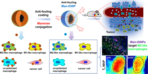
Although tumor-associated macrophages (TAMs) have been shown to promote cancer progression, their roles in tumor development and resistance to therapy remain to be fully understood, mainly because of the lack of a good approach to evaluate the dynamic changes of heterogeneous macrophages in their residing microenvironment. Here, we report an approach of using antibiofouling PEG-b-AGE polymer-coated iron oxide nanoparticles (IONPs) for targeted imaging of mannose receptor (CD206)-expressing M2-like TAMs. Antibiofouling polymer coatings block non-specific phagocytosis of IONPs by different cells but enable ligand-mediated CD206+ M2-like macrophage targeting after surface functionalizing with mannose (Man-IONP). Costaining tissue sections of the 4T1 mouse mammary tumors using an anti-CD206 antibody and fluorescent dye (TRITC)-labeled Man-IONP showed 94.7 ± 4.5% colocalization of TRITC-Man-IONPs with the anti-CD206 antibody. At 48 h after intravenous (i.v.) injection of Man-IONPs, magnetic resonance imaging of mice bearing orthotopic 4T1 mammary tumors showed a significantly larger IONP-induced decrease of the transverse relaxation time (T2) in tumors with 29.4 ± 1.5 ms compared to 12.3 ± 3.6 ms in tumors that received non-targeted IONP probes (P < 0.001). Immunofluorescence-stained tumor tissue sections collected at 6, 18, and 24 h after i.v. administration of the nanoprobes revealed that Man-IONPs specifically targeted CD206+ M2-like macrophages in various tumor areas at all time points, while nonconjugated IONPs were absent in the tumor after 18 h. Thus, antibiofouling Man-IONPs demonstrated the capability of explicitly imaging CD206+ M2-like macrophages in vivo and potentials for investigating the dynamics of macrophages in the tumor microenvironment and delivering therapeutics targeting M2-like TAMs.
中文翻译:

使用甘露糖共轭抗生物污损磁性氧化铁纳米颗粒对表达 CD206 的肿瘤相关 M2 样巨噬细胞进行靶向成像
尽管肿瘤相关巨噬细胞 (TAM) 已被证明可促进癌症进展,但它们在肿瘤发展和治疗抵抗中的作用仍有待充分了解,主要是因为缺乏一种评估其体内异质巨噬细胞动态变化的好方法。居住微环境。在这里,我们报告了一种使用抗生物污染 PEG- b -AGE 聚合物包覆的氧化铁纳米粒子 (IONP) 对表达甘露糖受体 (CD206) 的 M2 样 TAM 进行靶向成像的方法。抗生物污损聚合物涂层可阻止不同细胞对 IONP 的非特异性吞噬作用,但可实现配体介导的 CD206 +用甘露糖 (Man-IONP) 表面功能化后的 M2 样巨噬细胞靶向。使用抗 CD206 抗体和荧光染料 (TRITC) 标记的 Man-IONP 对 4T1 小鼠乳腺肿瘤的组织切片进行染色显示,TRITC-Man-IONP 与抗 CD206 抗体的共定位率为 94.7 ± 4.5%。在静脉 (iv) 注射 Man-IONP 后 48 小时,携带原位 4T1 乳腺肿瘤的小鼠的磁共振成像显示,IONP 诱导的肿瘤横向弛豫时间 ( T 2 )显着降低,与 29.4 ± 1.5 ms 相比,在接受非靶向 IONP 探针的肿瘤中为 12.3 ± 3.6 ms ( P< 0.001)。在静脉注射纳米探针后 6、18 和 24 小时收集的免疫荧光染色的肿瘤组织切片显示,Man-IONPs在所有时间点都特异性靶向不同肿瘤区域的CD206 + M2 样巨噬细胞,而未结合的 IONPs 在所有时间点均不存在。 18 小时后的肿瘤。因此,抗生物污染 Man-IONP 证明了在体内对CD206 + M2 样巨噬细胞进行显式成像的能力,以及研究肿瘤微环境中巨噬细胞动力学和提供靶向 M2 样 TAM 的治疗方法的潜力。
更新日期:2020-07-20
中文翻译:
使用甘露糖共轭抗生物污损磁性氧化铁纳米颗粒对表达 CD206 的肿瘤相关 M2 样巨噬细胞进行靶向成像
尽管肿瘤相关巨噬细胞 (TAM) 已被证明可促进癌症进展,但它们在肿瘤发展和治疗抵抗中的作用仍有待充分了解,主要是因为缺乏一种评估其体内异质巨噬细胞动态变化的好方法。居住微环境。在这里,我们报告了一种使用抗生物污染 PEG- b -AGE 聚合物包覆的氧化铁纳米粒子 (IONP) 对表达甘露糖受体 (CD206) 的 M2 样 TAM 进行靶向成像的方法。抗生物污损聚合物涂层可阻止不同细胞对 IONP 的非特异性吞噬作用,但可实现配体介导的 CD206 +用甘露糖 (Man-IONP) 表面功能化后的 M2 样巨噬细胞靶向。使用抗 CD206 抗体和荧光染料 (TRITC) 标记的 Man-IONP 对 4T1 小鼠乳腺肿瘤的组织切片进行染色显示,TRITC-Man-IONP 与抗 CD206 抗体的共定位率为 94.7 ± 4.5%。在静脉 (iv) 注射 Man-IONP 后 48 小时,携带原位 4T1 乳腺肿瘤的小鼠的磁共振成像显示,IONP 诱导的肿瘤横向弛豫时间 ( T 2 )显着降低,与 29.4 ± 1.5 ms 相比,在接受非靶向 IONP 探针的肿瘤中为 12.3 ± 3.6 ms ( P< 0.001)。在静脉注射纳米探针后 6、18 和 24 小时收集的免疫荧光染色的肿瘤组织切片显示,Man-IONPs在所有时间点都特异性靶向不同肿瘤区域的CD206 + M2 样巨噬细胞,而未结合的 IONPs 在所有时间点均不存在。 18 小时后的肿瘤。因此,抗生物污染 Man-IONP 证明了在体内对CD206 + M2 样巨噬细胞进行显式成像的能力,以及研究肿瘤微环境中巨噬细胞动力学和提供靶向 M2 样 TAM 的治疗方法的潜力。


 京公网安备 11010802027423号