当前位置:
X-MOL 学术
›
J. Biophotonics
›
论文详情
Our official English website, www.x-mol.net, welcomes your feedback! (Note: you will need to create a separate account there.)
Quantification of diabetic macular ischemia using novel three-dimensional optical coherence tomography angiography metrics.
Journal of Biophotonics ( IF 2.8 ) Pub Date : 2020-06-11 , DOI: 10.1002/jbio.202000152 Enrico Borrelli 1 , Riccardo Sacconi 1 , Lea Querques 1 , Marco Battista 1 , Francesco Bandello 1 , Giuseppe Querques 1
Journal of Biophotonics ( IF 2.8 ) Pub Date : 2020-06-11 , DOI: 10.1002/jbio.202000152 Enrico Borrelli 1 , Riccardo Sacconi 1 , Lea Querques 1 , Marco Battista 1 , Francesco Bandello 1 , Giuseppe Querques 1
Affiliation
|
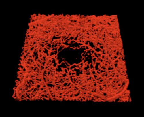
We applied three‐dimensional (3D) analysis to optical coherence tomography angiography (OCTA) to measure macular ischemia in eyes affected by non‐proliferative diabetic retinopathy (DR). A previously validated algorithm was applied to OCTA data in order to obtain 3D visualization of the retinal vasculature. Successively, a global thresholding algorithm was applied and two novel quantitative metrics were introduced: 3D vascular volume and 3D perfusion density. Two‐dimensional (2D) OCTA metrics were also obtained with different binarization thresholds for comparison. Of the 30 patients included, 15 were diagnosed with DR and 15 were controls. The 3D vascular volume and 3D perfusion density were reduced in DR eyes (P < .0001). The 2D variables also significantly differ between groups. The 3D perfusion density had the highest area under the receiver operating characteristic curve (0.964) among tested variables. Assessing quantitative perfusion using 3D analysis is reliable and promising, and with an elevated diagnostic efficacy in identifying DR eyes.
中文翻译:

定量使用新型三维光学相干断层扫描血管造影指标的糖尿病性黄斑缺血。
我们将三维(3D)分析应用于光学相干断层扫描血管造影(OCTA),以测量受非增生性糖尿病视网膜病变(DR)影响的眼睛的黄斑缺血。先前经过验证的算法已应用于OCTA数据,以获得视网膜脉管系统的3D可视化。随后,应用了全局阈值算法,并引入了两个新颖的定量指标:3D血管体积和3D灌注密度。还获得了具有不同二值化阈值的二维(2D)OCTA度量标准,以进行比较。在纳入的30位患者中,有15位被诊断患有DR,15位是对照组。DR眼睛的3D血管体积和3D灌注密度降低(P<.0001)。两组之间的2D变量也显着不同。在测试变量中,3D灌注密度在接收器工作特性曲线下(0.964)最高。使用3D分析评估定量灌注是可靠且有前途的,并且在识别DR眼睛方面具有提高的诊断功效。
更新日期:2020-06-11
中文翻译:
定量使用新型三维光学相干断层扫描血管造影指标的糖尿病性黄斑缺血。
我们将三维(3D)分析应用于光学相干断层扫描血管造影(OCTA),以测量受非增生性糖尿病视网膜病变(DR)影响的眼睛的黄斑缺血。先前经过验证的算法已应用于OCTA数据,以获得视网膜脉管系统的3D可视化。随后,应用了全局阈值算法,并引入了两个新颖的定量指标:3D血管体积和3D灌注密度。还获得了具有不同二值化阈值的二维(2D)OCTA度量标准,以进行比较。在纳入的30位患者中,有15位被诊断患有DR,15位是对照组。DR眼睛的3D血管体积和3D灌注密度降低(P<.0001)。两组之间的2D变量也显着不同。在测试变量中,3D灌注密度在接收器工作特性曲线下(0.964)最高。使用3D分析评估定量灌注是可靠且有前途的,并且在识别DR眼睛方面具有提高的诊断功效。


 京公网安备 11010802027423号