Current Bioinformatics ( IF 4 ) Pub Date : 2020-02-29 , DOI: 10.2174/1574893614666190723115832 Xiaogen Zhou 1 , Zuoyong Li 2 , Huosheng Xie 1 , Ting Feng 1 , Yan Lu 1 , Chuansheng Wang 3 , Rongyan Chen 4
|
Aims: The proposed method falls into the category of medical image processing.
Background: Computer-aided automatic analysis systems for the analysis and cytometry of leukocyte (White Blood Cells, WBCs) in human blood smear images are a powerful diagnostic tool for many types of diseases, such as anemia, malaria, syphilis, heavy metal poisoning, and leukemia. Leukocyte segmentation is a basis of its automatic analysis, and the segmentation accuracy will directly influence the reliability of image-based automatic leukocyte analysis.
Objective: This paper aims to present a leukocyte segmentation method, which improves segmentation accuracy under rapid and standard staining conditions.
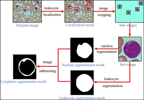
Methods: The proposed method first localizes leukocytes by color component combination and Adaptive Histogram Thresholding (AHT), and crops sub-image corresponding to each leukocyte. Then, the proposed method employs AHT to extract the nucleus of leukocyte and utilizes image color features to remove image backgrounds such as red blood cells and dyeing impurities. Finally, Canny edge detection is performed to extract the entire leukocyte. Accordingly, cytoplasm is obtained by subtracting nucleus with leukocyte.
Results: Experimental results on two datasets containing 160 leukocyte images show that the proposed method obtains more accurate segmentation results than their counterparts.
Conclusion: The proposed method obtains more accurate segmentation results than their counterparts under rapid and standard staining conditions.
中文翻译:
基于自适应直方图阈值和轮廓检测的白细胞图像分割
目的:提出的方法属于医学图像处理领域。
背景:计算机辅助自动分析系统可对人血涂片图像中的白细胞(白细胞,白细胞)进行分析和细胞计数,是针对多种疾病(如贫血,疟疾,梅毒,重金属中毒,和白血病。白细胞分割是其自动分析的基础,而分割的准确性将直接影响基于图像的自动白细胞分析的可靠性。
目的:本文旨在提出一种白细胞分割方法,该方法可提高快速和标准染色条件下的分割精度。
方法:所提出的方法首先通过颜色成分组合和自适应直方图阈值(AHT)定位白细胞,并裁剪与每个白细胞相对应的子图像。然后,提出的方法采用AHT提取白细胞核,并利用图像颜色特征去除图像背景,例如红细胞和染色杂质。最后,执行Canny边缘检测以提取整个白细胞。因此,通过用白细胞减去细胞核获得细胞质。
结果:在两个包含160个白细胞图像的数据集上的实验结果表明,所提出的方法比其对应方法可获得更准确的分割结果。
结论:在快速和标准染色条件下,该方法获得的分割结果比其对应方法更为准确。


 京公网安备 11010802027423号