当前位置:
X-MOL 学术
›
ACS Chem. Neurosci.
›
论文详情
Our official English website, www.x-mol.net, welcomes your feedback! (Note: you will need to create a separate account there.)
Glymphatic System Visualized by Chemical-Exchange-Saturation-Transfer Magnetic Resonance Imaging.
ACS Chemical Neuroscience ( IF 5 ) Pub Date : 2020-06-03 , DOI: 10.1021/acschemneuro.0c00222 Yuanfeng Chen 1 , Zhuozhi Dai 1 , Ruhang Fan 2 , David John Mikulis 3 , Jinming Qiu 1 , Zhiwei Shen 1 , Runrun Wang 1 , Lihua Lai 4 , Yanyan Tang 1 , Yan Li 1 , Yanlong Jia 1 , Gen Yan 5 , Renhua Wu 1, 6
ACS Chemical Neuroscience ( IF 5 ) Pub Date : 2020-06-03 , DOI: 10.1021/acschemneuro.0c00222 Yuanfeng Chen 1 , Zhuozhi Dai 1 , Ruhang Fan 2 , David John Mikulis 3 , Jinming Qiu 1 , Zhiwei Shen 1 , Runrun Wang 1 , Lihua Lai 4 , Yanyan Tang 1 , Yan Li 1 , Yanlong Jia 1 , Gen Yan 5 , Renhua Wu 1, 6
Affiliation

|
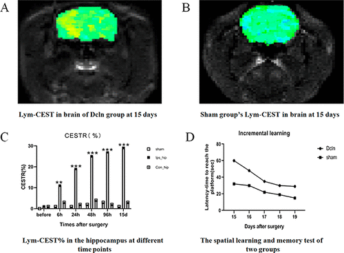
Dysfunction of the glymphatic system may play a significant role in the development of neurodegenerative diseases. However, in vivo imaging of the glymphatic system is challenging. In this study, we describe an unconventional MRI method for imaging the glymphatic system based on chemical exchange saturation transfer, which we tested in an in vivo porcine model of impaired glymphatic function. The blood, lymph, and cerebrospinal fluid (CSF) from one pig were used for testing the MRI effect in vitro at 7 Tesla (T). Unilateral deep cervical lymph node ligation models were then performed in 20 adult male Sprague–Dawley rats. The brains were scanned in vivo dynamically after surgery using the new MRI method. Behavioral tests were performed after each scanning session and the results were tested for correlations with the MRI signal intensity. Finally, the pathological assessment was conducted in the same brain slices. The special MRI effect in the lymph was evident at about 1.0 ppm in water and was distinguishable from those of blood and CSF. In the model group, the intensity of this MRI signal was significantly higher in the ipsilateral than in the contralateral hippocampus. The correlation between the signal abnormality and the behavioral score was significant (Pearson’s, R2 = 0.9154, p < 0.005). We conclude that the novel MRI method can visualize the glymphatic system in vivo.
中文翻译:

通过化学交换-饱和-转移磁共振成像可视化的淋巴系统。
淋巴系统的功能障碍可能在神经退行性疾病的发展中起重要作用。然而,体内对淋巴系统的成像是具有挑战性的。在这项研究中,我们描述了一种基于化学交换饱和转移的非常规MRI成像方法,用于淋巴系统成像,我们在受损的淋巴功能受损的体内猪模型中进行了测试。用一只猪的血液,淋巴液和脑脊髓液(CSF)在7特斯拉(T)处进行MRI体外测试。然后在20只成年雄性Sprague-Dawley大鼠中进行单侧深颈淋巴结结扎模型。体内扫描了大脑使用新的MRI方法动态地在手术后。在每次扫描后进行行为测试,并测试结果与MRI信号强度的相关性。最后,在相同的脑切片中进行病理评估。在水中约1.0 ppm时,对淋巴管的特殊MRI效果很明显,并且与血液和CSF有明显区别。在模型组中,同侧海马的MRI信号强度明显高于对侧海马。信号异常与行为评分之间的相关性很显着(Pearson's,R 2 = 0.9154,p <0.005)。我们得出结论,新颖的MRI方法可以可视化体内的淋巴系统。
更新日期:2020-07-01
中文翻译:

通过化学交换-饱和-转移磁共振成像可视化的淋巴系统。
淋巴系统的功能障碍可能在神经退行性疾病的发展中起重要作用。然而,体内对淋巴系统的成像是具有挑战性的。在这项研究中,我们描述了一种基于化学交换饱和转移的非常规MRI成像方法,用于淋巴系统成像,我们在受损的淋巴功能受损的体内猪模型中进行了测试。用一只猪的血液,淋巴液和脑脊髓液(CSF)在7特斯拉(T)处进行MRI体外测试。然后在20只成年雄性Sprague-Dawley大鼠中进行单侧深颈淋巴结结扎模型。体内扫描了大脑使用新的MRI方法动态地在手术后。在每次扫描后进行行为测试,并测试结果与MRI信号强度的相关性。最后,在相同的脑切片中进行病理评估。在水中约1.0 ppm时,对淋巴管的特殊MRI效果很明显,并且与血液和CSF有明显区别。在模型组中,同侧海马的MRI信号强度明显高于对侧海马。信号异常与行为评分之间的相关性很显着(Pearson's,R 2 = 0.9154,p <0.005)。我们得出结论,新颖的MRI方法可以可视化体内的淋巴系统。


 京公网安备 11010802027423号