Molecular Therapy - Methods & Clinical Development ( IF 4.7 ) Pub Date : 2020-06-04 , DOI: 10.1016/j.omtm.2020.06.001 Yongzheng Li 1, 2 , Jing Zhang 2, 3 , Zhenxuan Cheng 4 , Ying Wang 2, 5 , Tingben Huang 1, 2 , Kaichen Lai 1, 2 , Xue Du 1, 2 , Zhiwei Jiang 1, 2 , Guoli Yang 1, 2
|
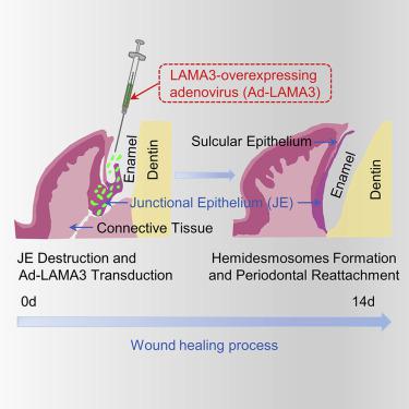
A robust dento-epithelial junction prevents external pathogenic factors from entering connective tissue and could be crucial for periodontal reattachment after periodontal surgery. The junctional epithelium (JE) is attached to the tooth surface through the hemidesmosome (HD) and internal basal lamina, where the primary component is laminin-332. Destruction of the JE leads to the loss of periodontal attachment. Traditional treatments are effective in eliminating local inflammation of the gingiva; however, few directly promote periodontal reattachment and HD formation. Here, we designed a gene-therapy strategy using the adenovirus-mediated human laminin-332 α3 chain (LAMA3) gene (Ad-LAMA3) transduced into a human-immortalized epidermal cell line (HaCaT) to study the formation of HD in vitro. Ad-LAMA3 promoted early adhesion and fast migration of HaCaT cells and increased expression of LAMA3 and type XVII collagen (BP180) significantly. Furthermore, HaCaT cells could facilitate formation of mature HDs after LAMA3 overexpression. In vivo experiments demonstrated that the JE transduced with Ad-LAMA3 could increase expression of LAMA3 and BP180 and “biological sealing” between the tooth and gingival epithelium. These results suggested that adenovirus-mediated LAMA3 transduction is a novel therapeutic strategy that promotes the stability and integration of the JE around the tooth during wound healing.
中文翻译:
腺病毒介导的LAMA3转导可增强伤口愈合过程中的血球小体形成和牙周附着。
牢固的牙本质-上皮连接可防止外部病原体进入结缔组织,并且对于牙周手术后的牙周再连接可能至关重要。交界上皮(JE)通过半球体(HD)和内部基底层附着在牙齿表面,主要成分是层粘连蛋白332。JE的破坏导致牙周附着的丧失。传统疗法可有效消除牙龈的局部炎症;但是,很少有直接促进牙周附着和HD形成的物质。在这里,我们设计了一种使用腺病毒介导的人类层粘连蛋白-332α3链(LAMA3)基因(Ad-LAMA3)进行基因治疗的策略,将其转导到人类永生化表皮细胞系(HaCaT)中,以研究体外高清的形成。Ad-LAMA3促进HaCaT细胞的早期粘附和快速迁移,并显着增加LAMA3和XVII型胶原蛋白(BP180)的表达。此外,HaCaT细胞可以促进LAMA3过表达后成熟HD的形成。体内实验表明,用Ad-LAMA3转导的JE可以增加LAMA3和BP180的表达以及牙齿与牙龈上皮之间的“生物密封”。这些结果表明,腺病毒介导的LAMA3转导是一种新型的治疗策略,可促进伤口愈合期间牙齿周围JE的稳定性和整合。


 京公网安备 11010802027423号