Gastrointestinal Endoscopy ( IF 7.7 ) Pub Date : 2020-06-04 , DOI: 10.1016/j.gie.2020.05.043 Hiromu Fukuda 1 , Ryu Ishihara 1 , Yusuke Kato 2 , Takashi Matsunaga 3 , Tsutomu Nishida 4 , Takuya Yamada 5 , Hideharu Ogiyama 6 , Mai Horie 7 , Kazuo Kinoshita 8 , Tomohiro Tada 9
|
Background and Aims
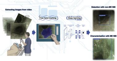
Narrow-band imaging (NBI) is currently regarded as the standard modality for diagnosing esophageal squamous cell carcinoma (SCC). We developed a computerized image-analysis system for diagnosing esophageal SCC by NBI and estimated its performance with video images.
Methods
Altogether, 23,746 images from 1544 pathologically proven superficial esophageal SCCs and 4587 images from 458 noncancerous and normal tissue were used to construct an artificial intelligence (AI) system. Five- to 9-second video clips from 144 patients captured by NBI or blue-light imaging were used as the validation dataset. These video images were diagnosed by the AI system and 13 board-certified specialists (experts).
Results
The diagnostic process was divided into 2 parts: detection (identify suspicious lesions) and characterization (differentiate cancer from noncancer). The sensitivities, specificities, and accuracies for the detection of SCC were, respectively, 91%, 51%, and 63% for the AI system and 79%, 72%, and 75% for the experts. The sensitivity of the AI system was significantly higher than that of the experts, but its specificity was significantly lower. Sensitivities, specificities, and accuracy for the characterization of SCC were, respectively, 86%, 89%, and 88% for the AI system and 74%, 76%, and 75% for the experts. The receiver operating characteristic curve showed that the AI system had significantly better diagnostic performance than the experts.
Conclusions
Our AI system showed significantly higher sensitivity for detecting SCC and higher accuracy for characterizing SCC from noncancerous tissue than endoscopic experts.
中文翻译:
人工智能与专家内镜医师在实时辅助诊断食管鳞状细胞癌中的性能比较(带视频)。
背景和目标
窄带成像(NBI)当前被认为是诊断食道鳞状细胞癌(SCC)的标准方法。我们开发了一种计算机图像分析系统,用于通过NBI诊断食管SCC,并通过视频图像评估其性能。
方法
总共使用了1544个经过病理证实的食管浅表食管鳞癌的23,746张图像和来自458个非癌组织和正常组织的4587张图像,以构建人工智能(AI)系统。通过NBI或蓝光成像捕获的144位患者的5至9秒视频剪辑用作验证数据集。这些视频图像是由AI系统和13位董事会认证的专家(专家)诊断出来的。
结果
诊断过程分为两个部分:检测(识别可疑病变)和表征(区分非癌性癌症)。对于AI系统,检测SCC的灵敏度,特异性和准确性分别为91%,51%和63%,对专家而言为79%,72%和75%。AI系统的灵敏度显着高于专家系统,但其特异性却明显较低。表征SCC的敏感性,特异性和准确性对于AI系统分别为86%,89%和88%,对专家而言分别为74%,76%和75%。接收机的工作特性曲线表明,人工智能系统比专家系统具有更好的诊断性能。
结论
与内窥镜专家相比,我们的AI系统显示出更高的检测SCC敏感性和表征非癌性组织SCC的准确性。


 京公网安备 11010802027423号