Biochimica et Biophysica Acta (BBA) - Molecular and Cell Biology of Lipids ( IF 4.8 ) Pub Date : 2020-06-03 , DOI: 10.1016/j.bbalip.2020.158753 Maciej Roman 1 , Tomasz P Wrobel 1 , Agnieszka Panek 1 , Czeslawa Paluszkiewicz 1 , Wojciech M Kwiatek 1
|
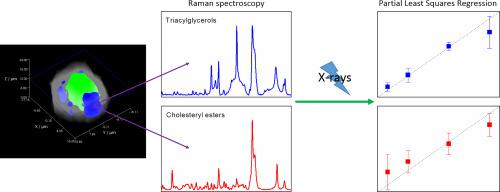
Lipid droplets (LDs) are key organelles in cancer cells proliferation, growth, and response to stress. These nanometric structures can aggregate to reach the size of microns becoming important cell components. Although it is known that LDs contain various lipids, their chemical composition is still under investigation. Moreover, their function in cell's response to exogenous factors is also not fully understood. Raman spectroscopy, together with chemometrics, has been shown to be a powerful tool for analytical analyses of cancer cell components on the subcellular level. It provides the opportunity to analyse LDs in a label-free manner in live cells. In the current study, this method was applied to investigate LDs composition in untreated and irradiated with X-ray beams prostate cancer cells. Raman mapping technique proved lipids accumulation in PC-3 cells and allowed visualization of LDs spatial distribution in cytoplasm. A heterogeneous composition of LDs was revealed by detailed analysis of Raman spectra. Interestingly, PC-3 cells were found to accumulate either triacylglycerols or cholesteryl esters. Finally, effect of X-ray radiation on the cells was investigated using Raman spectroscopy and fluorescence staining. Significant influence of LDs in the process of cell response was confirmed and time dependence of this phenomenon was determined.
中文翻译:
通过拉曼光谱研究了前列腺癌细胞中的脂质液滴和辐射的影响。
脂滴(LDs)是癌细胞增殖,生长和对压力的反应中的关键细胞器。这些纳米结构可以聚集达到微米的大小,成为重要的细胞成分。尽管已知LDs包含各种脂质,但其化学组成仍在研究中。而且,它们在细胞对外源因子的应答中的功能也没有被完全理解。拉曼光谱法和化学计量学已被证明是在亚细胞水平上分析癌细胞成分的强大工具。它提供了以无标记方式在活细胞中分析LD的机会。在当前的研究中,此方法用于研究未经治疗和用X射线束治疗的前列腺癌细胞的LD组成。拉曼作图技术证明了脂质在PC-3细胞中的积累,并使LDs在细胞质中的空间分布可视化。通过拉曼光谱的详细分析揭示了LD的异质组成。有趣的是,发现PC-3细胞会积聚三酰基甘油或胆固醇酯。最后,使用拉曼光谱和荧光染色研究了X射线辐射对细胞的影响。LDs在细胞反应过程中的重要影响得到确认,并确定了这种现象的时间依赖性。使用拉曼光谱和荧光染色研究了X射线辐射对细胞的影响。LDs在细胞反应过程中的重要影响得到确认,并确定了这种现象的时间依赖性。使用拉曼光谱和荧光染色研究了X射线辐射对细胞的影响。LDs在细胞反应过程中的重要影响得到确认,并确定了这种现象的时间依赖性。


 京公网安备 11010802027423号