当前位置:
X-MOL 学术
›
J. Anal. At. Spectrom.
›
论文详情
Our official English website, www.x-mol.net, welcomes your feedback! (Note: you will need to create a separate account there.)
Proof-of-concept for 2D/CT element analysis of entire cryofrozen islets of Langerhans using a cryoloop synchrotron X-ray fluorescence setup
Journal of Analytical Atomic Spectrometry ( IF 3.4 ) Pub Date : 2020-06-02 , DOI: 10.1039/d0ja00067a Björn De Samber 1, 2, 3, 4, 5 , Mohammed Bensellam 6, 7, 8, 9, 10 , Stijn J. M. Van Malderen 1, 2, 3, 4, 5 , Frank Seiboth 11, 12, 13 , Dennis Brückner 11, 12, 13, 14, 15 , Jan Garrevoet 11, 12, 13 , Gerald Falkenberg 11, 12, 13 , Jean-Christophe Jonas 6, 7, 8, 9, 10 , Laszlo Vincze 1, 2, 3, 4, 5
Journal of Analytical Atomic Spectrometry ( IF 3.4 ) Pub Date : 2020-06-02 , DOI: 10.1039/d0ja00067a Björn De Samber 1, 2, 3, 4, 5 , Mohammed Bensellam 6, 7, 8, 9, 10 , Stijn J. M. Van Malderen 1, 2, 3, 4, 5 , Frank Seiboth 11, 12, 13 , Dennis Brückner 11, 12, 13, 14, 15 , Jan Garrevoet 11, 12, 13 , Gerald Falkenberg 11, 12, 13 , Jean-Christophe Jonas 6, 7, 8, 9, 10 , Laszlo Vincze 1, 2, 3, 4, 5
Affiliation
|
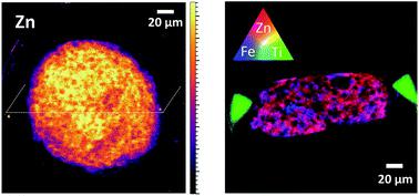
This work reports on synchrotron X-ray fluorescence (SR-XRF) imaging of vitrified islets of Langerhans in two-dimensional and computed tomography (CT) mode and serves as a proof-of-principle for the preparation and SR-XRF imaging of cryoloops in a cryogenic environment. Selection of suited cryoloops and cryoprotectant solution enabled vitrification of the islets. The SR-XRF experimental setup was adapted with a cryogenic gas stream, enabling analysis of cryofrozen islets and a dual silicon drift detector (SDD) configuration, providing an increased solid angle for XRF collection. Element distributions of K, Ca, Fe and Zn within single, cryofrozen islets could be obtained with 500 nm spatial resolution using X-ray compound refractive lenses (CRLs) with an adaptive phase plate. Titanium, present as a trace element in the cryoloops, was used as a marker signal to efficiently define the XRF scan area. Preliminary 2D cryo-analysis of wild type (n = 1) and Mt1-Mt2-knock-out (n = 1) islets under low/high glucose conditions did not reveal changes in the distribution and concentration of Ca, Fe and Zn. Cryotomography XRF scans indicated aspherical islets within the cryoloop, likely due to capillary effects in the cryoloop before plunge freezing. The presence of diffracting ice crystals hampered CT reconstruction, indicating the need for further development of a routine workflow ensuring perfect vitrification. Collectively, this novel multidisciplinary approach proved to be valid and can be used for future studies to assess elemental compositions of islets of Langerhans or other cell clusters/tissues under different pathophysiological conditions.
中文翻译:

使用冷冻回路同步加速器X射线荧光装置对朗格汉斯的整个冷冻冷冻胰岛进行2D / CT元素分析的概念验证
这项工作报告了二维和计算机断层扫描(CT)模式下朗格汉斯玻璃化胰岛的同步加速器X射线荧光(SR-XRF)成像,并为冷冻环的制备和SR-XRF成像提供了原理证明在低温环境中。选择合适的冷冻环和冷冻保护剂溶液可使胰岛玻璃化。SR-XRF实验装置适用于低温气流,可以分析冷冻的胰岛和双硅漂移检测器(SDD)配置,从而为XRF收集提供更大的立体角。使用具有自适应相位板的X射线复合折射透镜(CRL),可以在500 nm空间分辨率下获得单个冷冻冷冻胰岛中K,Ca,Fe和Zn的元素分布。钛作为冷冻回路中的微量元素存在,用作标记信号以有效定义XRF扫描区域。野生型的初步二维冷冻分析(n = 1)和Mt1-Mt2-敲除胰岛(n = 1)在低/高葡萄糖条件下的胰岛未揭示Ca,Fe和Zn的分布和浓度的变化。冷冻X射线摄影(XRF)扫描表明,冷冻回路内的非球面岛很可能是由于急冻前冷冻回路中的毛细管效应所致。衍射冰晶的存在阻碍了CT的重建,表明需要进一步开发确保完美玻璃化的常规工作流程。总的来说,这种新颖的多学科方法被证明是有效的,可用于将来的研究,以评估在不同病理生理条件下朗格汉斯岛或其他细胞簇/组织的胰岛的元素组成。
更新日期:2020-07-08
中文翻译:
使用冷冻回路同步加速器X射线荧光装置对朗格汉斯的整个冷冻冷冻胰岛进行2D / CT元素分析的概念验证
这项工作报告了二维和计算机断层扫描(CT)模式下朗格汉斯玻璃化胰岛的同步加速器X射线荧光(SR-XRF)成像,并为冷冻环的制备和SR-XRF成像提供了原理证明在低温环境中。选择合适的冷冻环和冷冻保护剂溶液可使胰岛玻璃化。SR-XRF实验装置适用于低温气流,可以分析冷冻的胰岛和双硅漂移检测器(SDD)配置,从而为XRF收集提供更大的立体角。使用具有自适应相位板的X射线复合折射透镜(CRL),可以在500 nm空间分辨率下获得单个冷冻冷冻胰岛中K,Ca,Fe和Zn的元素分布。钛作为冷冻回路中的微量元素存在,用作标记信号以有效定义XRF扫描区域。野生型的初步二维冷冻分析(n = 1)和Mt1-Mt2-敲除胰岛(n = 1)在低/高葡萄糖条件下的胰岛未揭示Ca,Fe和Zn的分布和浓度的变化。冷冻X射线摄影(XRF)扫描表明,冷冻回路内的非球面岛很可能是由于急冻前冷冻回路中的毛细管效应所致。衍射冰晶的存在阻碍了CT的重建,表明需要进一步开发确保完美玻璃化的常规工作流程。总的来说,这种新颖的多学科方法被证明是有效的,可用于将来的研究,以评估在不同病理生理条件下朗格汉斯岛或其他细胞簇/组织的胰岛的元素组成。


 京公网安备 11010802027423号