当前位置:
X-MOL 学术
›
J. Anal. At. Spectrom.
›
论文详情
Our official English website, www.x-mol.net, welcomes your feedback! (Note: you will need to create a separate account there.)
Development of X-ray imaging of intracellular elements and structure
Journal of Analytical Atomic Spectrometry ( IF 3.4 ) Pub Date : 2020-05-29 , DOI: 10.1039/d0ja00128g Satoshi Matsuyama 1, 2, 3, 4, 5 , Kazuhiro Maeshima 5, 6, 7, 8, 9 , Mari Shimura 5, 6, 10, 11, 12
Journal of Analytical Atomic Spectrometry ( IF 3.4 ) Pub Date : 2020-05-29 , DOI: 10.1039/d0ja00128g Satoshi Matsuyama 1, 2, 3, 4, 5 , Kazuhiro Maeshima 5, 6, 7, 8, 9 , Mari Shimura 5, 6, 10, 11, 12
Affiliation
|
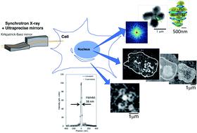
The desire to see the smallest possible objects, such as the contents of cells, reflects our intellectual curiosity and has resulted in the development of various types of microscopes. Microscopes using an X-ray source were developed after Röntgen's discovery of X-rays in 1895. Röntgen rays were first used for photography in 1896 and for observation of the structural details of biological samples in the 1900s. This use of X-rays grew considerably following the development of X-ray optics such as diffractive lenses and total-reflection mirrors in the late 1940s. X-ray microscopy theoretically has better resolution than that of visible light (400–700 nm) microscopy and has developed differently from both visible light and electron microscopy due to the penetration ability of X-rays. The third-generation synchrotron radiation facilities that produce higher electron beam energies promoted X-ray microprobes for various types of microscopies. The accompanying development of X-ray focusing systems has led to today's submicron X-ray probes, which have high enough resolution for imaging cells at the organelle level. In this review, we describe the imaging technologies using synchrotron X-ray fluorescence by means of a sub-100 nm focusing system and X-ray diffraction, which facilitates the determination of the cellular elemental distribution and structure.
中文翻译:

细胞内元素和结构的X射线成像技术发展
希望看到最小的物体(例如细胞的内容)反映了我们的知识好奇心,并导致了各种类型显微镜的发展。1895年伦琴(Röntgen)发现X射线之后,便开发了使用X射线源的显微镜。伦琴射线(Röntgen)射线于1896年首次用于摄影,并于1900年代用于观察生物样品的结构细节。在1940年代后期,诸如衍射透镜和全反射镜之类的X射线光学器件的发展之后,对X射线的使用大大增加。X射线显微镜在理论上比可见光(400-700 nm)显微镜具有更好的分辨率,并且由于X射线的穿透能力,其发展与可见光和电子显微镜不同。产生更高电子束能量的第三代同步加速器辐射设施促进了各种类型显微镜的X射线微探针。X射线聚焦系统的伴随发展导致了当今的亚微米X射线探针,该探针具有足够高的分辨率,可以在细胞器水平对细胞进行成像。在这篇综述中,我们描述了通过低于100 nm的聚焦系统和X射线衍射使用同步加速器X射线荧光的成像技术,这有助于确定细胞的元素分布和结构。
更新日期:2020-07-08
中文翻译:
细胞内元素和结构的X射线成像技术发展
希望看到最小的物体(例如细胞的内容)反映了我们的知识好奇心,并导致了各种类型显微镜的发展。1895年伦琴(Röntgen)发现X射线之后,便开发了使用X射线源的显微镜。伦琴射线(Röntgen)射线于1896年首次用于摄影,并于1900年代用于观察生物样品的结构细节。在1940年代后期,诸如衍射透镜和全反射镜之类的X射线光学器件的发展之后,对X射线的使用大大增加。X射线显微镜在理论上比可见光(400-700 nm)显微镜具有更好的分辨率,并且由于X射线的穿透能力,其发展与可见光和电子显微镜不同。产生更高电子束能量的第三代同步加速器辐射设施促进了各种类型显微镜的X射线微探针。X射线聚焦系统的伴随发展导致了当今的亚微米X射线探针,该探针具有足够高的分辨率,可以在细胞器水平对细胞进行成像。在这篇综述中,我们描述了通过低于100 nm的聚焦系统和X射线衍射使用同步加速器X射线荧光的成像技术,这有助于确定细胞的元素分布和结构。


 京公网安备 11010802027423号