当前位置:
X-MOL 学术
›
J. Biophotonics
›
论文详情
Our official English website, www.x-mol.net, welcomes your feedback! (Note: you will need to create a separate account there.)
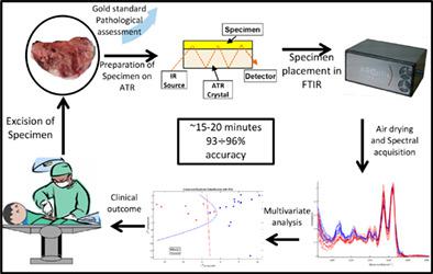
Rapid intraoperative diagnosis of gynecological cancer by ATR-FTIR spectroscopy of fresh tissue biopsy.
Journal of Biophotonics ( IF 2.8 ) Pub Date : 2020-06-23 , DOI: 10.1002/jbio.202000114 Dov Malonek 1 , Ben-Zion Dekel 1 , Gabi Haran 2, 3 , Renat Reens-Carmel 2, 3 , Gabriel M Groisman 3, 4 , Mordechai Hallak 2, 3 , Ilan Bruchim 2, 3
Journal of Biophotonics ( IF 2.8 ) Pub Date : 2020-06-23 , DOI: 10.1002/jbio.202000114 Dov Malonek 1 , Ben-Zion Dekel 1 , Gabi Haran 2, 3 , Renat Reens-Carmel 2, 3 , Gabriel M Groisman 3, 4 , Mordechai Hallak 2, 3 , Ilan Bruchim 2, 3
Affiliation
|
A rapid and reliable intraoperative diagnostic technique to support clinical decisions was developed using Fourier‐transform infrared (FTIR) spectroscopy. Twenty‐six fresh tissue samples were collected intraoperatively from patients undergoing gynecological surgeries. Frozen section (FS) histopathology aimed to discriminate between malignant and benign tumors was performed, and attenuated total reflection (ATR) FTIR spectra were collected from these samples. Digital dehydration and principal component analysis and linear discriminant analysis (PCA‐LDA) models were developed to classify samples into malignant and benign groups. Two validation schemes were employed: k‐fold and “leave one out.” FTIR absorption spectrum of a fresh tissue sample was obtained in less than 5 minutes. The fingerprint spectral region of malignant tumors was consistently different from that of benign tumors. The PCA‐LDA discrimination model correctly classified the samples into malignant and benign groups with accuracies of 96% and 93% for the k‐fold and “leave one out” validation schemes, respectively. We showed that a simple tissue preparation followed by ATR‐FTIR spectroscopy provides accurate means for very rapid tumor classification into malignant and benign gynecological tumors. With further development, the proposed method has high potential to be used as an adjunct to the intraoperative FS histopathology technique.
中文翻译:

通过新鲜组织活检的ATR-FTIR光谱术对妇科癌症进行术中快速诊断。
使用傅里叶变换红外(FTIR)光谱技术开发了一种快速可靠的术中诊断技术来支持临床决策。术中从妇科手术患者中收集了26份新鲜组织样本。进行旨在区分恶性和良性肿瘤的冰冻切片(FS)组织病理学,并从这些样品中收集了衰减全反射(ATR)FTIR光谱。开发了数字脱水,主成分分析和线性判别分析(PCA-LDA)模型,以将样品分为恶性和良性组。采用了两种验证方案:k折叠和“遗漏”。在不到5分钟的时间内获得了新鲜组织样品的FTIR吸收光谱。恶性肿瘤的指纹光谱区与良性肿瘤的指纹区一致。PCA-LDA辨别模型将样本正确分类为恶性组和良性组,其k倍验证方案和“遗漏”验证方案的准确率分别为96%和93%。我们证明了简单的组织准备,然后进行ATR-FTIR光谱分析,为将肿瘤快速分类为恶性和良性妇科肿瘤提供了准确的方法。随着进一步的发展,该方法具有较高的潜力,可作为术中FS组织病理学技术的辅助手段。分别。我们证明了简单的组织准备,然后进行ATR-FTIR光谱分析,为将肿瘤快速分类为恶性和良性妇科肿瘤提供了准确的方法。随着进一步的发展,该方法具有较高的潜力,可作为术中FS组织病理学技术的辅助手段。分别。我们证明了简单的组织准备,然后进行ATR-FTIR光谱分析,为将肿瘤快速分类为恶性和良性妇科肿瘤提供了准确的方法。随着进一步的发展,该方法具有较高的潜力,可作为术中FS组织病理学技术的辅助手段。
更新日期:2020-06-23
中文翻译:
通过新鲜组织活检的ATR-FTIR光谱术对妇科癌症进行术中快速诊断。
使用傅里叶变换红外(FTIR)光谱技术开发了一种快速可靠的术中诊断技术来支持临床决策。术中从妇科手术患者中收集了26份新鲜组织样本。进行旨在区分恶性和良性肿瘤的冰冻切片(FS)组织病理学,并从这些样品中收集了衰减全反射(ATR)FTIR光谱。开发了数字脱水,主成分分析和线性判别分析(PCA-LDA)模型,以将样品分为恶性和良性组。采用了两种验证方案:k折叠和“遗漏”。在不到5分钟的时间内获得了新鲜组织样品的FTIR吸收光谱。恶性肿瘤的指纹光谱区与良性肿瘤的指纹区一致。PCA-LDA辨别模型将样本正确分类为恶性组和良性组,其k倍验证方案和“遗漏”验证方案的准确率分别为96%和93%。我们证明了简单的组织准备,然后进行ATR-FTIR光谱分析,为将肿瘤快速分类为恶性和良性妇科肿瘤提供了准确的方法。随着进一步的发展,该方法具有较高的潜力,可作为术中FS组织病理学技术的辅助手段。分别。我们证明了简单的组织准备,然后进行ATR-FTIR光谱分析,为将肿瘤快速分类为恶性和良性妇科肿瘤提供了准确的方法。随着进一步的发展,该方法具有较高的潜力,可作为术中FS组织病理学技术的辅助手段。分别。我们证明了简单的组织准备,然后进行ATR-FTIR光谱分析,为将肿瘤快速分类为恶性和良性妇科肿瘤提供了准确的方法。随着进一步的发展,该方法具有较高的潜力,可作为术中FS组织病理学技术的辅助手段。


 京公网安备 11010802027423号