The Journal of Thoracic and Cardiovascular Surgery ( IF 6 ) Pub Date : 2020-05-28 , DOI: 10.1016/j.jtcvs.2020.04.169 Fangqiu Fu 1 , Yang Zhang 1 , Shengping Wang 2 , Yuan Li 3 , Zezhou Wang 4 , Hong Hu 1 , Haiquan Chen 1
|
Objective
Pure ground-glass nodules are considered to be radiologically noninvasive in lung adenocarcinoma. However, some pure ground-glass nodules are found to be invasive adenocarcinoma pathologically. This study aims to identify the computed tomography parameters distinguishing invasive adenocarcinoma from adenocarcinoma in situ and minimally invasive adenocarcinoma.
Methods
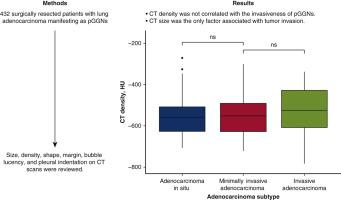
From May 2011 to December 2015, patients with completely resected adenocarcinoma appearing as pure ground-glass nodules were reviewed. To evaluate the association between computed tomography features and the invasiveness of pure ground-glass nodules, logistic regression analyses were conducted.
Results
Among 432 enrolled patients, 118 (27.3%) were classified as adenocarcinoma in situ, 213 (49.3%) were classified as minimally invasive adenocarcinoma, 101 (23.4%) were classified as invasive adenocarcinoma. There was no postoperative recurrence for patients with pure ground-glass nodules. Logistic regression analyses demonstrated that computed tomography size was the only independent radiographic factor associated with adenocarcinoma in situ (odds ratio, 47.165; 95% confidence interval, 19.279-115.390; P < .001), whereas computed tomography density was not (odds ratio, 1.002; 95% confidence interval, 0.999-1.005; P = .127). Further analyses revealed that there was no distributional difference in computed tomography density among 3 groups (P = .173). Even after propensity score matching for adenocarcinoma in situ/minimally invasive adenocarcinoma and invasive adenocarcinoma, no significant difference in computed tomography density was observed (P = .741). The subanalyses for pure ground-glass nodules with 1 cm or more in size also indicated similar results.
Conclusions
In patients with pure ground-glass nodules, computed tomography size was the only radiographic parameter associated with tumor invasion. Measuring computed tomography density provided no advantage in differentiating invasive adenocarcinoma from adenocarcinoma in situ and minimally invasive adenocarcinoma.
中文翻译:
计算机断层扫描密度与纯磨玻璃结节的病理性肿瘤侵袭无关
客观的
在肺腺癌中,纯磨玻璃结节被认为是放射学上无创的。然而,一些纯磨玻璃结节在病理上被发现是浸润性腺癌。本研究旨在确定区分浸润性腺癌与原位腺癌和微浸润性腺癌的计算机断层扫描参数。
方法
2011 年 5 月至 2015 年 12 月,对完全切除的腺癌表现为纯磨玻璃结节的患者进行回顾性分析。为了评估计算机断层扫描特征与纯磨玻璃结节侵袭性之间的关联,进行了逻辑回归分析。
结果
432例入组患者中,原位腺癌118例(27.3%),微浸润腺癌213例(49.3%),浸润性腺癌101例(23.4%)。纯磨玻璃结节患者术后无复发。Logistic 回归分析表明,计算机断层扫描大小是与原位腺癌相关的唯一独立影像学因素(优势比,47.165;95% 置信区间,19.279-115.390;P < .001),而计算机断层扫描密度不是(优势比, 1.002;95% 置信区间,0.999-1.005;P = .127 )。进一步分析显示,3组间计算机断层扫描密度没有分布差异(P = .173)。即使在对原位腺癌/微浸润腺癌和浸润性腺癌进行倾向评分匹配后,也没有观察到计算机断层扫描密度的显着差异 ( P = .741)。对大小为 1 cm 或更大的纯磨玻璃结节的亚组分析也表明了类似的结果。
结论
在纯磨玻璃结节患者中,计算机断层扫描大小是与肿瘤侵袭相关的唯一影像学参数。测量计算机断层扫描密度在区分浸润性腺癌与原位腺癌和微浸润性腺癌方面没有优势。


 京公网安备 11010802027423号