当前位置:
X-MOL 学术
›
J. Comp. Neurol.
›
论文详情
Our official English website, www.x-mol.net, welcomes your feedback! (Note: you will need to create a separate account there.)
Nectin-2α is localized at cholinergic neuron dendrites and regulates synapse formation in the medial habenula.
The Journal of Comparative Neurology ( IF 2.5 ) Pub Date : 2020-05-26 , DOI: 10.1002/cne.24958 Hajime Shiotani 1 , Muneaki Miyata 1 , Takeshi Kameyama 1 , Kenji Mandai 1, 2, 3 , Miwako Yamasaki 4 , Masahiko Watanabe 4 , Kiyohito Mizutani 1 , Yoshimi Takai 1
The Journal of Comparative Neurology ( IF 2.5 ) Pub Date : 2020-05-26 , DOI: 10.1002/cne.24958 Hajime Shiotani 1 , Muneaki Miyata 1 , Takeshi Kameyama 1 , Kenji Mandai 1, 2, 3 , Miwako Yamasaki 4 , Masahiko Watanabe 4 , Kiyohito Mizutani 1 , Yoshimi Takai 1
Affiliation
|
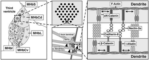
The medial habenula (MHb) receives afferents from the triangular septum and the medial septal complex, projects efferents to the interpeduncular nucleus (IPN) in the midbrain to regulate dopamine and serotonin levels, and is implicated in stress, depression, memory, and nicotine withdrawal syndrome. We previously showed that the cell adhesion molecule nectin‐2α is localized at the boundary between adjacent somata of clustered cholinergic neurons and regulates the voltage‐gated A‐type K+ channel Kv4.2 localization at membrane specializations in the MHb. This adhesion apparatus, named nectin‐2α spots, is not associated with the nectin‐binding protein afadin or any classic cadherins and their binding proteins p120‐catenin and β‐catenin. We showed here that nectin‐2α was additionally localized at cholinergic neuron dendrites in synaptic regions of the MHb. The genetic ablation of nectin‐2 reduced the number of synapses in the MHb without affecting their morphology. Nectin‐2α was associated with afadin, cadherin‐8, p120‐catenin, β‐catenin, and αN‐catenin, forming puncta adherentia junctions (PAJs). Nectin‐2α was observed in the IPN, but not in the triangular septum or the medial septal complex. The genetic ablation of nectin‐2 did not affect synapse formation in the IPN. These results indicate that nectin‐2α forms two types of adhesion apparatus in the MHb, namely nectin‐2α spots at neighboring somata and PAJs at neighboring dendrites, and that dendritic PAJs regulate synapse formation in the MHb.
中文翻译:

Nectin-2α 位于胆碱能神经元树突,调节内侧缰核中的突触形成。
内侧缰核 (MHb) 接收来自三角隔和内侧隔复合体的传入神经,将传出神经投射到中脑的脚间核 (IPN) 以调节多巴胺和血清素水平,并与压力、抑郁、记忆和尼古丁戒断有关综合征。我们之前表明,细胞粘附分子 nectin-2α 位于成簇的胆碱能神经元的相邻胞体之间的边界处,并调节电压门控 A 型 K +通道 Kv4.2 定位在 MHb 中的膜特化处。这种称为 nectin-2α 斑点的粘附装置与 nectin 结合蛋白 afadin 或任何经典的钙粘蛋白及其结合蛋白 p120-catenin 和 β-catenin 无关。我们在这里展示了 nectin-2α 还定位于 MHb 突触区域的胆碱能神经元树突。nectin-2的遗传消融减少了 MHb 中突触的数量,而不影响它们的形态。Nectin-2α 与 afadin、cadherin-8、p120-catenin、β-catenin 和 αN-catenin 相关,形成点状粘附连接(PAJs)。在 IPN 中观察到 Nectin-2α,但在三角隔或内侧隔复合体中未观察到。nectin-2的基因消融不影响 IPN 中的突触形成。这些结果表明 nectin-2α 在 MHb 中形成两种类型的粘附装置,即相邻胞体的 nectin-2α 点和相邻树突的 PAJ,树突 PAJ 调节 MHb 中的突触形成。
更新日期:2020-06-29
中文翻译:
Nectin-2α 位于胆碱能神经元树突,调节内侧缰核中的突触形成。
内侧缰核 (MHb) 接收来自三角隔和内侧隔复合体的传入神经,将传出神经投射到中脑的脚间核 (IPN) 以调节多巴胺和血清素水平,并与压力、抑郁、记忆和尼古丁戒断有关综合征。我们之前表明,细胞粘附分子 nectin-2α 位于成簇的胆碱能神经元的相邻胞体之间的边界处,并调节电压门控 A 型 K +通道 Kv4.2 定位在 MHb 中的膜特化处。这种称为 nectin-2α 斑点的粘附装置与 nectin 结合蛋白 afadin 或任何经典的钙粘蛋白及其结合蛋白 p120-catenin 和 β-catenin 无关。我们在这里展示了 nectin-2α 还定位于 MHb 突触区域的胆碱能神经元树突。nectin-2的遗传消融减少了 MHb 中突触的数量,而不影响它们的形态。Nectin-2α 与 afadin、cadherin-8、p120-catenin、β-catenin 和 αN-catenin 相关,形成点状粘附连接(PAJs)。在 IPN 中观察到 Nectin-2α,但在三角隔或内侧隔复合体中未观察到。nectin-2的基因消融不影响 IPN 中的突触形成。这些结果表明 nectin-2α 在 MHb 中形成两种类型的粘附装置,即相邻胞体的 nectin-2α 点和相邻树突的 PAJ,树突 PAJ 调节 MHb 中的突触形成。


 京公网安备 11010802027423号