当前位置:
X-MOL 学术
›
J. Biophotonics
›
论文详情
Our official English website, www.x-mol.net, welcomes your feedback! (Note: you will need to create a separate account there.)
High-speed X-ray-induced luminescence computed tomography.
Journal of Biophotonics ( IF 2.8 ) Pub Date : 2020-06-23 , DOI: 10.1002/jbio.202000066 Xianjin Dai 1 , Kai Cheng 1 , Wei Zhao 1 , Lei Xing 1
Journal of Biophotonics ( IF 2.8 ) Pub Date : 2020-06-23 , DOI: 10.1002/jbio.202000066 Xianjin Dai 1 , Kai Cheng 1 , Wei Zhao 1 , Lei Xing 1
Affiliation
|
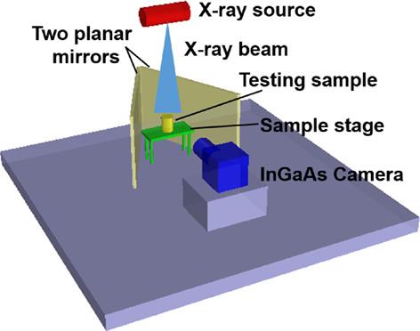
X‐ray‐induced luminescence computed tomography (XLCT) is an emerging molecular imaging. Challenges in improving spatial resolution and reducing the scan time in a whole‐body field of view (FOV) still remain for practical in vivo applications. In this study, we present a novel XLCT technique capable of obtaining three‐dimensional (3D) images from a single snapshot. Specifically, a customed two‐planar‐mirror component is integrated into a cone beam XLCT imaging system to obtain multiple optical views of an object simultaneously. Furthermore, a compressive sensing based algorithm is adopted to improve the efficiency of 3D XLCT image reconstruction. Numerical simulations and experiments were conducted to validate the single snapshot X‐ray‐induced luminescence computed tomography (SS‐XLCT). The results show that the 3D distribution of the nanophosphor targets can be visualized much faster than conventional cone beam XLCT imaging method that was used in our comparisons while maintaining comparable spatial resolution as in conventional XLCT imaging. SS‐XLCT has the potential to harness the power of XLCT for rapid whole‐body in vivo molecular imaging of small animals.
中文翻译:

高速 X 射线诱导发光计算机断层扫描。
X射线诱导发光计算机断层扫描(XLCT)是一种新兴的分子成像。对于体内实际应用来说,提高空间分辨率和减少全身视场(FOV)扫描时间仍然面临挑战。在这项研究中,我们提出了一种新颖的 XLCT 技术,能够从单个快照中获取三维 (3D) 图像。具体来说,将定制的双平面镜组件集成到锥束 XLCT 成像系统中,以同时获得物体的多个光学视图。此外,采用基于压缩感知的算法来提高3D XLCT图像重建的效率。进行数值模拟和实验以验证单快照 X 射线诱导发光计算机断层扫描 (SS-XLCT)。结果表明,与我们比较中使用的传统锥束 XLCT 成像方法相比,纳米磷光体目标的 3D 分布可视化速度要快得多,同时保持与传统 XLCT 成像相当的空间分辨率。SS-XLCT 有潜力利用 XLCT 的力量对小动物进行快速全身体内分子成像。
更新日期:2020-06-23
中文翻译:
高速 X 射线诱导发光计算机断层扫描。
X射线诱导发光计算机断层扫描(XLCT)是一种新兴的分子成像。对于体内实际应用来说,提高空间分辨率和减少全身视场(FOV)扫描时间仍然面临挑战。在这项研究中,我们提出了一种新颖的 XLCT 技术,能够从单个快照中获取三维 (3D) 图像。具体来说,将定制的双平面镜组件集成到锥束 XLCT 成像系统中,以同时获得物体的多个光学视图。此外,采用基于压缩感知的算法来提高3D XLCT图像重建的效率。进行数值模拟和实验以验证单快照 X 射线诱导发光计算机断层扫描 (SS-XLCT)。结果表明,与我们比较中使用的传统锥束 XLCT 成像方法相比,纳米磷光体目标的 3D 分布可视化速度要快得多,同时保持与传统 XLCT 成像相当的空间分辨率。SS-XLCT 有潜力利用 XLCT 的力量对小动物进行快速全身体内分子成像。


 京公网安备 11010802027423号