当前位置:
X-MOL 学术
›
ACS Biomater. Sci. Eng.
›
论文详情
Our official English website, www.x-mol.net, welcomes your feedback! (Note: you will need to create a separate account there.)
Rapid Prototyping of Multilayer Microphysiological Systems
ACS Biomaterials Science & Engineering ( IF 5.8 ) Pub Date : 2020-05-20 , DOI: 10.1021/acsbiomaterials.0c00190 Sanjin Hosic 1 , Adam J Bindas 1 , Marissa L Puzan 1 , Will Lake 1 , Jonathan R Soucy 1 , Fanny Zhou 2 , Ryan A Koppes 1 , David T Breault 2, 3, 4 , Shashi K Murthy 1 , Abigail N Koppes 1, 5
ACS Biomaterials Science & Engineering ( IF 5.8 ) Pub Date : 2020-05-20 , DOI: 10.1021/acsbiomaterials.0c00190 Sanjin Hosic 1 , Adam J Bindas 1 , Marissa L Puzan 1 , Will Lake 1 , Jonathan R Soucy 1 , Fanny Zhou 2 , Ryan A Koppes 1 , David T Breault 2, 3, 4 , Shashi K Murthy 1 , Abigail N Koppes 1, 5
Affiliation
|
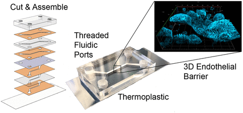
Microfluidic organs-on-chips aim to realize more biorelevant in vitro experiments compared to traditional two-dimensional (2D) static cell culture. Often such devices are fabricated via poly(dimethylsiloxane) (PDMS) soft lithography, which offers benefits (e.g., high feature resolution) along with drawbacks (e.g., prototyping time/costs). Here, we report benchtop fabrication of multilayer, PDMS-free, thermoplastic organs-on-chips via laser cut and assembly with double-sided adhesives that overcome some limitations of traditional PDMS lithography. Cut and assembled chips are economical to prototype ($2 per chip), can be fabricated in parallel within hours, and are Luer compatible. Biocompatibility was demonstrated with epithelial line Caco-2 cells and primary human small intestinal organoids. Comparable to control static Transwell cultures, Caco-2 and organoids cultured on chips formed confluent monolayers expressing tight junctions with low permeability. Caco-2 cells-on-chip differentiated ∼4 times faster, including increased mucus, compared to controls. To demonstrate the robustness of cut and assemble, we fabricated a dual membrane, trilayer chip integrating 2D and 3D compartments with accessible apical and basolateral flow chambers. As proof of concept, we cocultured a human, differentiated monolayer and intact 3D organoids within multilayered contacting compartments. The epithelium exhibited 3D tissue structure and organoids expanded close to the adjacent monolayer, retaining proliferative stem cells over 10 days. Taken together, cut and assemble offers the capability to rapidly and economically manufacture microfluidic devices, thereby presenting a compelling fabrication technique for developing organs-on-chips of various geometries to study multicellular tissues.
中文翻译:

多层微生理系统的快速原型设计
与传统的二维 (2D) 静态细胞培养相比,微流体器官芯片旨在实现更多生物相关的体外实验。通常这样的设备是通过聚(二甲基硅氧烷)(PDMS)软光刻制造的,它提供了好处(例如,高特征分辨率)以及缺点(例如,原型制作时间/成本)。在这里,我们报告了通过激光切割和组装双面粘合剂在台式制造多层、不含 PDMS 的热塑性片上器官,克服了传统 PDMS 光刻的一些局限性。切割和组装的芯片对于原型来说是经济的(每个芯片 2 美元),可以在几小时内并行制造,并且与 Luer 兼容。上皮细胞系 Caco-2 细胞和原代人类小肠类器官证明了生物相容性。与控制静态 Transwell 培养物相比,在芯片上培养的 Caco-2 和类器官形成融合的单层,表达低渗透性的紧密连接。与对照相比,Caco-2 芯片上的细胞分化速度提高了约 4 倍,包括粘液增加。为了证明切割和组装的稳健性,我们制造了一个双膜、三层芯片,将 2D 和 3D 隔室与可访问的顶端和基底侧流室集成在一起。作为概念证明,我们在多层接触隔间内共培养了一个人类、分化的单层和完整的 3D 类器官。上皮表现出 3D 组织结构,类器官在邻近单层附近扩张,保留增殖干细胞超过 10 天。总之,切割和组装提供了快速、经济地制造微流体设备的能力,
更新日期:2020-05-20
中文翻译:
多层微生理系统的快速原型设计
与传统的二维 (2D) 静态细胞培养相比,微流体器官芯片旨在实现更多生物相关的体外实验。通常这样的设备是通过聚(二甲基硅氧烷)(PDMS)软光刻制造的,它提供了好处(例如,高特征分辨率)以及缺点(例如,原型制作时间/成本)。在这里,我们报告了通过激光切割和组装双面粘合剂在台式制造多层、不含 PDMS 的热塑性片上器官,克服了传统 PDMS 光刻的一些局限性。切割和组装的芯片对于原型来说是经济的(每个芯片 2 美元),可以在几小时内并行制造,并且与 Luer 兼容。上皮细胞系 Caco-2 细胞和原代人类小肠类器官证明了生物相容性。与控制静态 Transwell 培养物相比,在芯片上培养的 Caco-2 和类器官形成融合的单层,表达低渗透性的紧密连接。与对照相比,Caco-2 芯片上的细胞分化速度提高了约 4 倍,包括粘液增加。为了证明切割和组装的稳健性,我们制造了一个双膜、三层芯片,将 2D 和 3D 隔室与可访问的顶端和基底侧流室集成在一起。作为概念证明,我们在多层接触隔间内共培养了一个人类、分化的单层和完整的 3D 类器官。上皮表现出 3D 组织结构,类器官在邻近单层附近扩张,保留增殖干细胞超过 10 天。总之,切割和组装提供了快速、经济地制造微流体设备的能力,


 京公网安备 11010802027423号