Our official English website, www.x-mol.net, welcomes your feedback! (Note: you will need to create a separate account there.)
An integrated pipeline for high-throughput screening and profiling of spheroids using simple live image analysis of frame to frame variations
Methods ( IF 4.8 ) Pub Date : 2020-05-01 , DOI: 10.1016/j.ymeth.2020.05.017 Haneen Alsehli 1 , Fuad Mosis 2 , Christopher Thompson 3 , Eva Hamrud 4 , Erika Wiseman 5 , Eileen Gentleman 6 , Davide Danovi 7
Methods ( IF 4.8 ) Pub Date : 2020-05-01 , DOI: 10.1016/j.ymeth.2020.05.017 Haneen Alsehli 1 , Fuad Mosis 2 , Christopher Thompson 3 , Eva Hamrud 4 , Erika Wiseman 5 , Eileen Gentleman 6 , Davide Danovi 7
Affiliation
|
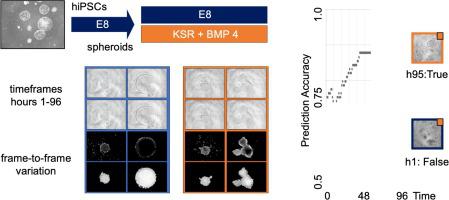
High throughput imaging methods can be applied to relevant cell culture models, fostering their use in research and translational applications. Improvements in microscopy, computational capabilities and data analysis have enabled high-throughput, high-content approaches from endpoint 2D microscopy images. Nonetheless, trade-offs in acquisition, computation and storage between content and throughput remain, in particular when cells and cell structures are imaged in 3D. Moreover, live 3D phase contrast microscopy images are not often amenable to analysis because of the high level of background noise. Cultures of Human induced pluripotent stem cells (hiPSC) offer unprecedented scope to profile and screen conditions affecting cell fate decisions, self-organisation and early embryonic development. However, quantifying changes in the morphology or function of cell structures derived from hiPSCs over time presents significant challenges. Here, we report a novel method based on the analysis of live phase contrast microscopy images of hiPSC spheroids. We compare self-renewing versus differentiating media conditions, which give rise to spheroids with distinct morphologies; round versus branched, respectively. These cell structures are segmented from 2D projections and analysed based on frame-to-frame variations. Importantly, a tailored convolutional neural network is trained and applied to predict culture conditions from time-frame images. We compare our results with more classic and involved endpoint 3D confocal microscopy and propose that such approaches can complement spheroid-based assays developed for the purpose of screening and profiling. This workflow can be realistically implemented in laboratories using imaging-based high-throughput methods for regenerative medicine and drug discovery.
中文翻译:

使用帧到帧变化的简单实时图像分析对球体进行高通量筛选和分析的集成管道
高通量成像方法可应用于相关细胞培养模型,促进其在研究和转化应用中的应用。显微技术、计算能力和数据分析方面的改进使端点二维显微图像的高通量、高内涵方法成为可能。尽管如此,内容和吞吐量之间的获取、计算和存储的权衡仍然存在,特别是当细胞和细胞结构以 3D 方式成像时。此外,由于背景噪声水平高,实时 3D 相差显微镜图像通常不适合分析。人类诱导多能干细胞 (hiPSC) 的培养物为分析和筛选影响细胞命运决定、自组织和早期胚胎发育的条件提供了前所未有的范围。然而,随着时间的推移,量化源自 hiPSC 的细胞结构的形态或功能变化是一项重大挑战。在这里,我们报告了一种基于 hiPSC 球体实时相差显微镜图像分析的新方法。我们比较了自我更新与分化的媒体条件,这会产生具有不同形态的球体;分别是圆形与分支。这些细胞结构是从 2D 投影中分割出来的,并基于帧到帧的变化进行分析。重要的是,定制的卷积神经网络经过训练并应用于从时间帧图像预测培养条件。我们将我们的结果与更经典和更复杂的端点 3D 共聚焦显微镜进行比较,并建议这种方法可以补充为筛选和分析目的而开发的基于球体的检测。
更新日期:2020-05-01
中文翻译:
使用帧到帧变化的简单实时图像分析对球体进行高通量筛选和分析的集成管道
高通量成像方法可应用于相关细胞培养模型,促进其在研究和转化应用中的应用。显微技术、计算能力和数据分析方面的改进使端点二维显微图像的高通量、高内涵方法成为可能。尽管如此,内容和吞吐量之间的获取、计算和存储的权衡仍然存在,特别是当细胞和细胞结构以 3D 方式成像时。此外,由于背景噪声水平高,实时 3D 相差显微镜图像通常不适合分析。人类诱导多能干细胞 (hiPSC) 的培养物为分析和筛选影响细胞命运决定、自组织和早期胚胎发育的条件提供了前所未有的范围。然而,随着时间的推移,量化源自 hiPSC 的细胞结构的形态或功能变化是一项重大挑战。在这里,我们报告了一种基于 hiPSC 球体实时相差显微镜图像分析的新方法。我们比较了自我更新与分化的媒体条件,这会产生具有不同形态的球体;分别是圆形与分支。这些细胞结构是从 2D 投影中分割出来的,并基于帧到帧的变化进行分析。重要的是,定制的卷积神经网络经过训练并应用于从时间帧图像预测培养条件。我们将我们的结果与更经典和更复杂的端点 3D 共聚焦显微镜进行比较,并建议这种方法可以补充为筛选和分析目的而开发的基于球体的检测。


 京公网安备 11010802027423号