当前位置:
X-MOL 学术
›
J. Struct. Biol.
›
论文详情
Our official English website, www.x-mol.net, welcomes your feedback! (Note: you will need to create a separate account there.)
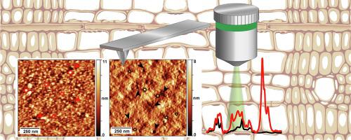
Atomic force microscopy imaging of delignified secondary cell walls in liquid conditions facilitates interpretation of wood ultrastructure.
Journal of Structural Biology ( IF 3 ) Pub Date : 2020-05-20 , DOI: 10.1016/j.jsb.2020.107532 Maria Adobes-Vidal 1 , Marion Frey 1 , Tobias Keplinger 1
Journal of Structural Biology ( IF 3 ) Pub Date : 2020-05-20 , DOI: 10.1016/j.jsb.2020.107532 Maria Adobes-Vidal 1 , Marion Frey 1 , Tobias Keplinger 1
Affiliation
|
Deep understanding of the physicochemical and structural characteristics of wood at the nanoscale is essential for improving wood usage in biorefining and advancing new high performance materials design. Herein, we use in situ atomic force microscopy and a simple delignification treatment to elucidate the nanoscale architecture of individual secondary cell wall layers. Advantages of this approach are: (i) minimal sample preparation that reduces the introduction of potential artifacts; (ii) prevention of structural rearrangements due to dehydration; (iii) increased accessibility to structural details masked by the lignin matrix; and (iv) possibility to complement results with other analytical techniques without sample manipulation. The methodology permits the visualization of parallel and helicoidally arranged microfibril aggregates in the S1 layer and the determination of lignin contribution to microfibril aggregates forming S2 layers. Cellulose and hemicelluloses constitute the core of the aggregates with a mean diameter of approximately 19 nm, and lignin encloses the core forming single structural entities of about 30 nm diameter. Furthermore, we highlight the implications of sample preparation and imaging parameters on the characterization of microfibril aggregates by AFM.
中文翻译:

在液体条件下脱木质素次生细胞壁的原子力显微镜成像有助于解释木材超微结构。
深入了解纳米级木材的物理化学和结构特征对于改善生物精炼中的木材使用和推进新的高性能材料设计至关重要。在此,我们使用原位原子力显微镜和简单的脱木素处理来阐明单个次生细胞壁层的纳米级结构。这种方法的优点是: (i) 最少的样品制备,减少了潜在伪影的引入;(ii) 防止因脱水引起的结构重排;(iii) 增加对被木质素基质掩盖的结构细节的可及性;(iv) 无需样品操作即可用其他分析技术补充结果的可能性。该方法允许可视化 S1 层中平行和螺旋排列的微纤维聚集体,并确定木质素对形成 S2 层的微纤维聚集体的贡献。纤维素和半纤维素构成了平均直径约为 19 nm 的聚集体的核心,木质素包围了核心,形成了直径约为 30 nm 的单个结构实体。此外,我们强调了样品制备和成像参数对 AFM 微纤维聚集体表征的影响。
更新日期:2020-05-20
中文翻译:
在液体条件下脱木质素次生细胞壁的原子力显微镜成像有助于解释木材超微结构。
深入了解纳米级木材的物理化学和结构特征对于改善生物精炼中的木材使用和推进新的高性能材料设计至关重要。在此,我们使用原位原子力显微镜和简单的脱木素处理来阐明单个次生细胞壁层的纳米级结构。这种方法的优点是: (i) 最少的样品制备,减少了潜在伪影的引入;(ii) 防止因脱水引起的结构重排;(iii) 增加对被木质素基质掩盖的结构细节的可及性;(iv) 无需样品操作即可用其他分析技术补充结果的可能性。该方法允许可视化 S1 层中平行和螺旋排列的微纤维聚集体,并确定木质素对形成 S2 层的微纤维聚集体的贡献。纤维素和半纤维素构成了平均直径约为 19 nm 的聚集体的核心,木质素包围了核心,形成了直径约为 30 nm 的单个结构实体。此外,我们强调了样品制备和成像参数对 AFM 微纤维聚集体表征的影响。


 京公网安备 11010802027423号