当前位置:
X-MOL 学术
›
Eur. J. Nerosci.
›
论文详情
Our official English website, www.x-mol.net, welcomes your feedback! (Note: you will need to create a separate account there.)
Early volumetric changes of hippocampus and medial prefrontal cortex following medial temporal lobe resection.
European Journal of Neroscience ( IF 3.4 ) Pub Date : 2020-05-18 , DOI: 10.1111/ejn.14784 Anna Pajkert 1 , Christoph J Ploner 1 , Thomas-Nicolas Lehmann 2 , Veronica A Witte 3 , Frank Oltmanns 4 , Werner Sommer 5 , Martin Holtkamp 1, 4 , Hauke R Heekeren 6, 7 , Carsten Finke 1, 8
European Journal of Neroscience ( IF 3.4 ) Pub Date : 2020-05-18 , DOI: 10.1111/ejn.14784 Anna Pajkert 1 , Christoph J Ploner 1 , Thomas-Nicolas Lehmann 2 , Veronica A Witte 3 , Frank Oltmanns 4 , Werner Sommer 5 , Martin Holtkamp 1, 4 , Hauke R Heekeren 6, 7 , Carsten Finke 1, 8
Affiliation
|
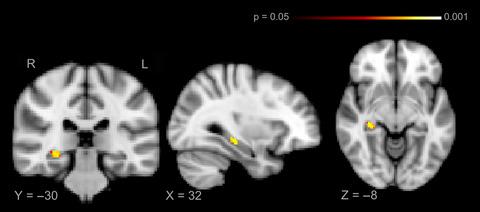
Previous studies have shown that cognitive demands and physical exercise stimulate adult neurogenesis in the dentate gyrus and hippocampus. Recent observations in healthy humans and patients with mild cognitive impairment moreover suggest that training‐induced increases in hippocampal volume may be associated with improved memory performance. The corresponding plasticity processes in hippocampal volume may occur on timescales of months to years. For patients with focal lesions in this region, previous functional imaging studies suggest that increased recruitment of the contralateral hippocampus and extratemporal regions may be an important part of the reorganization of episodic memory. However, it is currently unclear whether focal damage to the medial temporal lobe (MTL) induces gray matter (GM) volume changes in the intact contralateral hippocampus and in connected network regions on a shorter timescale. We therefore investigated whether unilateral resection of the MTL, including the hippocampus, induces measurable volumetric changes in the contralateral hippocampus and in the default mode network (DMN). We recruited 31 patients with unilateral left (N = 19) or right (N = 12) hippocampal sclerosis undergoing MTL resection for treatment of drug‐resistant epilepsy. Structural MRI was acquired immediately before and 3 months after surgery. Longitudinal voxel‐based morphometry (VBM) analysis revealed a significant increase of right hippocampal volume following resection of the left anterior MTL. Furthermore, this patient group showed GM volume increases in the DMN. These results demonstrate significant structural plasticity of the contralateral hippocampus, even in patients with a long‐standing unilateral hippocampal dysfunction and structural reorganization processes extending to distant, but functionally connected brain regions.
中文翻译:

内侧颞叶切除后海马和内侧前额叶皮层的早期体积变化。
先前的研究表明,认知需求和体育锻炼会刺激齿状回和海马中的成人神经发生。此外,在健康人和轻度认知障碍患者中的最新观察结果表明,训练导致的海马体容量增加可能与记忆力改善有关。海马体积的相应可塑性过程可能会发生数月至数年。对于在该区域有局灶性病变的患者,以前的功能影像学研究表明,对侧海马和颞外区域的募集增加可能是情节记忆重组的重要组成部分。然而,目前尚不清楚对内侧颞叶(MTL)的局灶性损伤是否会在较短的时间内在完整的对侧海马和连接的网络区域中引起灰质(GM)体积变化。因此,我们调查了单侧切除包括海马在内的MTL是否在对侧海马和默认模式网络(DMN)中引起可测量的体积变化。我们招募了31例单侧左(N = 19)或右(N = 12)接受MTL切除术治疗耐药性癫痫的海马硬化。术前和术后3个月进行结构性MRI检查。基于纵向体素的形态计量学(VBM)分析显示,切除左前MTL后右海马体积显着增加。此外,该患者组显示DMN中的GM量增加。这些结果表明对侧海马具有显着的结构可塑性,即使在长期存在单侧海马功能障碍且结构重组过程延伸至遥远但功能相连的大脑区域的患者中也是如此。
更新日期:2020-05-18
中文翻译:
内侧颞叶切除后海马和内侧前额叶皮层的早期体积变化。
先前的研究表明,认知需求和体育锻炼会刺激齿状回和海马中的成人神经发生。此外,在健康人和轻度认知障碍患者中的最新观察结果表明,训练导致的海马体容量增加可能与记忆力改善有关。海马体积的相应可塑性过程可能会发生数月至数年。对于在该区域有局灶性病变的患者,以前的功能影像学研究表明,对侧海马和颞外区域的募集增加可能是情节记忆重组的重要组成部分。然而,目前尚不清楚对内侧颞叶(MTL)的局灶性损伤是否会在较短的时间内在完整的对侧海马和连接的网络区域中引起灰质(GM)体积变化。因此,我们调查了单侧切除包括海马在内的MTL是否在对侧海马和默认模式网络(DMN)中引起可测量的体积变化。我们招募了31例单侧左(N = 19)或右(N = 12)接受MTL切除术治疗耐药性癫痫的海马硬化。术前和术后3个月进行结构性MRI检查。基于纵向体素的形态计量学(VBM)分析显示,切除左前MTL后右海马体积显着增加。此外,该患者组显示DMN中的GM量增加。这些结果表明对侧海马具有显着的结构可塑性,即使在长期存在单侧海马功能障碍且结构重组过程延伸至遥远但功能相连的大脑区域的患者中也是如此。


 京公网安备 11010802027423号