当前位置:
X-MOL 学术
›
J. Biophotonics
›
论文详情
Our official English website, www.x-mol.net, welcomes your feedback! (Note: you will need to create a separate account there.)
Computed tomography for in vivo deep over-1000 nm near-infrared fluorescence imaging.
Journal of Biophotonics ( IF 2.8 ) Pub Date : 2020-06-08 , DOI: 10.1002/jbio.202000071 Masakazu Umezawa 1 , Toshihiro Sera 2 , Hideo Yokota 3 , Maho Takematsu 1 , Masahiko Morita 3 , Gil Yeroslavsky 4 , Masao Kamimura 1, 4 , Kohei Soga 1, 4
Journal of Biophotonics ( IF 2.8 ) Pub Date : 2020-06-08 , DOI: 10.1002/jbio.202000071 Masakazu Umezawa 1 , Toshihiro Sera 2 , Hideo Yokota 3 , Maho Takematsu 1 , Masahiko Morita 3 , Gil Yeroslavsky 4 , Masao Kamimura 1, 4 , Kohei Soga 1, 4
Affiliation
|
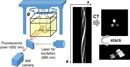
This study aims to develop a novel cross‐sectional imaging of fluorescence in over‐1000 nm near‐infrared (OTN‐NIR), which allows in vivo deep imaging, using computed tomography (CT) system. Cylindrical specimens of composite of OTN‐NIR fluorophore, NaGdF4 co‐doped with Yb3+ and Ho3+ (ex: 980 nm, em: 1150 nm), were embedded in cubic agar (10.5–12 mm) or in the peritoneal cavity of mice and placed on a rotatable stage. When the fluorescence from inside of the samples was serially captured from multiple angles, the images were disrupted by the reflection and refraction of emitted light on the sample‐air interface. Immersing the sample into water filled in a rectangular bath suppressed the disruption at the interface and successfully reconstructed the position and concentration of OTN‐NIR fluorophores on the cross‐sectional images using a CT technique. This is promising as a novel three‐dimensional imaging technique for OTN‐NIR fluorescent image projections of small animals captured from multiple angles.
中文翻译:

用于体内1000纳米以上深红外荧光成像的计算机断层扫描。
这项研究的目的是开发一种新颖的横截面荧光成像技术,该技术在超过1000 nm的近红外(OTN-NIR)中使用计算机断层扫描(CT)系统进行体内深层成像。OTN-NIR荧光团,掺有Yb 3+和Ho 3+的NaGdF 4复合材料的圆柱样品(例如:980 nm,em:1150 nm),被包埋在立方琼脂(10.5–12 mm)或小鼠的腹膜腔中,并置于可旋转平台上。当从多个角度连续捕获样品内部的荧光时,图像会由于样品-空气界面上发射光的反射和折射而被破坏。将样品浸入装在矩形槽中的水中,可以抑制界面处的破裂,并使用CT技术成功地重建了横截面图像上OTN-NIR荧光团的位置和浓度。这是一种新颖的三维成像技术,可用于从多个角度捕获的小动物的OTN-NIR荧光图像投影。
更新日期:2020-06-08
中文翻译:
用于体内1000纳米以上深红外荧光成像的计算机断层扫描。
这项研究的目的是开发一种新颖的横截面荧光成像技术,该技术在超过1000 nm的近红外(OTN-NIR)中使用计算机断层扫描(CT)系统进行体内深层成像。OTN-NIR荧光团,掺有Yb 3+和Ho 3+的NaGdF 4复合材料的圆柱样品(例如:980 nm,em:1150 nm),被包埋在立方琼脂(10.5–12 mm)或小鼠的腹膜腔中,并置于可旋转平台上。当从多个角度连续捕获样品内部的荧光时,图像会由于样品-空气界面上发射光的反射和折射而被破坏。将样品浸入装在矩形槽中的水中,可以抑制界面处的破裂,并使用CT技术成功地重建了横截面图像上OTN-NIR荧光团的位置和浓度。这是一种新颖的三维成像技术,可用于从多个角度捕获的小动物的OTN-NIR荧光图像投影。


 京公网安备 11010802027423号