IRBM ( IF 4.8 ) Pub Date : 2020-05-08 , DOI: 10.1016/j.irbm.2020.04.004 C. Caredda , L. Mahieu-Williame , R. Sablong , M. Sdika , J. Guyotat , B. Montcel
|
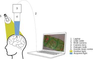
Intraoperative optical imaging is a localization technique for the functional areas of the human brain cortex during neurosurgical procedures. However, it still lacks robustness to be used as a clinical standard. In particular new biomarkers of brain functionality with improved sensitivity and specificity are needed. We present a method for the real time identification of the activated cortical areas based on the analysis of the cortical hemodynamic using a RGB camera and a white light source. We measure the quantitative oxy and deoxy-hemoglobin concentration changes in the human brain cortex with the modified Beer-Lambert law and Monte Carlo simulations. A functional model has been implemented to define in real time a binary biomarker of the cortical activation following neuronal activation by physiological stimuli. The results show a good correlation between the computed activation maps and the brain areas localized by electrical brain stimulation. We demonstrate that a RGB camera combined with a quantitative modeling of brain hemodynamics biomarkers can evaluate in real time the functional areas during neurosurgery and serve as a tool of choice to complement electrical brain stimulation.
中文翻译:
基于RGB成像的实时术中功能性脑图
术中光学成像是神经外科手术过程中人脑皮质功能区域的定位技术。但是,它仍然缺乏用作临床标准的鲁棒性。特别地,需要具有改善的敏感性和特异性的脑功能的新生物标志物。我们提出了一种基于RGB摄像头和白光源的皮质血流动力学分析为基础的实时识别激活的皮质区域的方法。我们用改良的比尔-朗伯定律和蒙特卡洛模拟法测量人脑皮质中的定量氧和脱氧血红蛋白浓度变化。已经实现了功能模型以实时定义在通过生理刺激的神经元激活之后皮层激活的二进制生物标记。结果表明,在计算出的激活图和脑电刺激定位的大脑区域之间有很好的相关性。我们证明了RGB相机与脑血流动力学生物标志物的定量建模相结合,可以实时评估神经外科手术期间的功能区域,并可以作为补充脑电刺激的选择工具。


 京公网安备 11010802027423号