当前位置:
X-MOL 学术
›
Hum. Brain Mapp.
›
论文详情
Our official English website, www.x-mol.net, welcomes your feedback! (Note: you will need to create a separate account there.)
In vivo characterization of emerging white matter microstructure in the fetal brain in the third trimester.
Human Brain Mapping ( IF 4.8 ) Pub Date : 2020-05-06 , DOI: 10.1002/hbm.25006 Camilo Jaimes 1, 2, 3 , Fedel Machado-Rivas 1, 3 , Onur Afacan 1, 3 , Shadab Khan 1, 3 , Bahram Marami 1, 3 , Cynthia M Ortinau 4 , Caitlin K Rollins 3, 5 , Clemente Velasco-Annis 1 , Simon K Warfield 1, 3 , Ali Gholipour 1, 3
Human Brain Mapping ( IF 4.8 ) Pub Date : 2020-05-06 , DOI: 10.1002/hbm.25006 Camilo Jaimes 1, 2, 3 , Fedel Machado-Rivas 1, 3 , Onur Afacan 1, 3 , Shadab Khan 1, 3 , Bahram Marami 1, 3 , Cynthia M Ortinau 4 , Caitlin K Rollins 3, 5 , Clemente Velasco-Annis 1 , Simon K Warfield 1, 3 , Ali Gholipour 1, 3
Affiliation
|
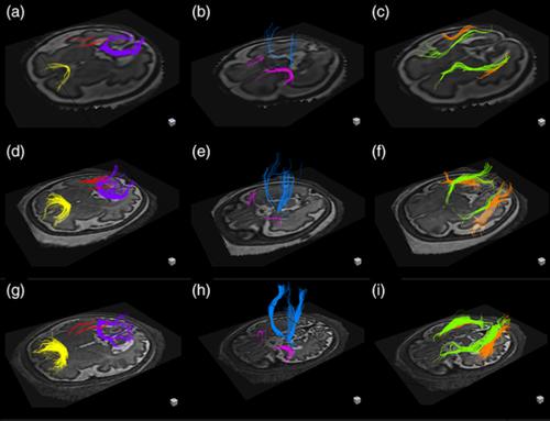
The third trimester of pregnancy is a period of rapid development of fiber bundles in the fetal white matter. Using a recently developed motion‐tracked slice‐to‐volume registration (MT‐SVR) method, we aimed to quantify tract‐specific developmental changes in apparent diffusion coefficient (ADC), fractional anisotropy (FA), and volume in third trimester healthy fetuses. To this end, we reconstructed diffusion tensor images from motion corrected fetal diffusion magnetic resonance imaging data. With an approved protocol, fetal MRI exams were performed on healthy pregnant women at 3 Tesla and included multiple (2–8) diffusion scans of the fetal head (1–2 b = 0 s/mm2 images and 12 diffusion‐sensitized images at b = 500 s/mm2). Diffusion data from 32 fetuses (13 females) with median gestational age (GA) of 33 weeks 4 days were processed with MT‐SVR and deterministic tractography seeded by regions of interest corresponding to 12 major fiber tracts. Multivariable regression analysis was used to evaluate the association of GA with volume, FA, and ADC for each tract. For all tracts, the volume and FA increased, and the ADC decreased with GA. Associations reached statistical significance for: FA and ADC of the forceps major; volume and ADC for the forceps minor; FA, ADC, and volume for the cingulum; ADC, FA, and volume for the uncinate fasciculi; ADC of the inferior fronto‐occipital fasciculi, ADC of the inferior longitudinal fasciculi; and FA and ADC for the corticospinal tracts. These quantitative results demonstrate the complex pattern and rates of tract‐specific, GA‐related microstructural changes of the developing white matter in human fetal brain.
中文翻译:

妊娠晚期胎儿大脑中新兴白质微结构的体内表征。
妊娠晚期是胎儿白质中纤维束快速发育的时期。使用最近开发的运动跟踪切片体积配准 (MT-SVR) 方法,我们旨在量化孕晚期健康胎儿表观扩散系数 (ADC)、各向异性分数 (FA) 和体积的特定道发育变化. 为此,我们从运动校正的胎儿扩散磁共振成像数据重建了扩散张量图像。根据批准的方案,在 3 特斯拉对健康孕妇进行胎儿 MRI 检查,包括多次 (2-8) 胎头弥散扫描(1-2 b = 0 s/mm 2图像和 12 b = 500 秒/毫米2)。来自 32 名胎儿(13 名女性)的中位孕龄(GA)为 33 周 4 天的扩散数据使用 MT-SVR 和确定性纤维束成像技术处理,这些数据由对应于 12 个主要纤维束的感兴趣区域播种。多变量回归分析用于评估 GA 与每个通道的体积、FA 和 ADC 的关联。对于所有区域,体积和 FA 增加,ADC 随 GA 降低。相关性达到统计显着性:镊子专业的FA和ADC;小镊子的体积和 ADC;FA、ADC 和扣带体积;ADC、FA 和钩状束的体积;额枕下束ADC、下纵束ADC;和 FA 和 ADC 用于皮质脊髓束。这些定量结果证明了特定区域的复杂模式和比率,
更新日期:2020-05-06
中文翻译:
妊娠晚期胎儿大脑中新兴白质微结构的体内表征。
妊娠晚期是胎儿白质中纤维束快速发育的时期。使用最近开发的运动跟踪切片体积配准 (MT-SVR) 方法,我们旨在量化孕晚期健康胎儿表观扩散系数 (ADC)、各向异性分数 (FA) 和体积的特定道发育变化. 为此,我们从运动校正的胎儿扩散磁共振成像数据重建了扩散张量图像。根据批准的方案,在 3 特斯拉对健康孕妇进行胎儿 MRI 检查,包括多次 (2-8) 胎头弥散扫描(1-2 b = 0 s/mm 2图像和 12 b = 500 秒/毫米2)。来自 32 名胎儿(13 名女性)的中位孕龄(GA)为 33 周 4 天的扩散数据使用 MT-SVR 和确定性纤维束成像技术处理,这些数据由对应于 12 个主要纤维束的感兴趣区域播种。多变量回归分析用于评估 GA 与每个通道的体积、FA 和 ADC 的关联。对于所有区域,体积和 FA 增加,ADC 随 GA 降低。相关性达到统计显着性:镊子专业的FA和ADC;小镊子的体积和 ADC;FA、ADC 和扣带体积;ADC、FA 和钩状束的体积;额枕下束ADC、下纵束ADC;和 FA 和 ADC 用于皮质脊髓束。这些定量结果证明了特定区域的复杂模式和比率,


 京公网安备 11010802027423号