当前位置:
X-MOL 学术
›
J. Struct. Biol.
›
论文详情
Our official English website, www.x-mol.net, welcomes your feedback! (Note: you will need to create a separate account there.)
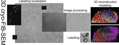
Cryo-FIB-SEM as a promising tool for localizing proteins in 3D.
Journal of Structural Biology ( IF 3 ) Pub Date : 2020-05-05 , DOI: 10.1016/j.jsb.2020.107528 Daniele Spehner 1 , Anna M Steyer 2 , Luca Bertinetti 3 , Igor Orlov 1 , Lucas Benoit 4 , Karin Pernet-Gallay 4 , Andreas Schertel 5 , Patrick Schultz 1
Journal of Structural Biology ( IF 3 ) Pub Date : 2020-05-05 , DOI: 10.1016/j.jsb.2020.107528 Daniele Spehner 1 , Anna M Steyer 2 , Luca Bertinetti 3 , Igor Orlov 1 , Lucas Benoit 4 , Karin Pernet-Gallay 4 , Andreas Schertel 5 , Patrick Schultz 1
Affiliation
|
Focused Ion Beam-Scanning Electron Microscopy (FIB-SEM) is an invaluable tool to visualize the 3D architecture of cell constituents and map cell networks. Recently, amorphous ice embedding techniques have been associated with FIB-SEM to ensure that the biological material remains as close as possible to its native state. Here we have vitrified human HeLa cells and directly imaged them by cryo-FIB-SEM with the secondary electron InLens detector at cryogenic temperature and without any staining. Image stacks were aligned and processed by denoising, removal of ion beam milling artefacts and local charge imbalance. Images were assembled into a 3D volume and the major cell constituents were modelled. The data illustrate the power of the workflow to provide a detailed view of the internal architecture of the fully hydrated, close-to-native, entire HeLa cell. In addition, we have studied the feasibility of combining cryo-FIB-SEM imaging with live-cell protein detection. We demonstrate that internalized gold particles can be visualized by detecting back scattered primary electrons at low kV while simultaneously acquiring signals from the secondary electron detector to image major cell features. Furthermore, gold-conjugated antibodies directed against RNA polymerase II could be observed in the endo-lysosomal pathway while labelling of the enzyme in the nucleus was not detected, a shortcoming likely due to the inadequacy between the size of the gold particles and the voxel size. With further refinements, this method promises to have a variety of applications where the goal is to localize cellular antigens while visualizing the entire native cell in three dimensions.
中文翻译:

Cryo-FIB-SEM 作为在 3D 中定位蛋白质的有前途的工具。
聚焦离子束扫描电子显微镜 (FIB-SEM) 是一种非常宝贵的工具,可用于可视化细胞成分的 3D 结构和映射细胞网络。最近,无定形冰嵌入技术已与 FIB-SEM 相关联,以确保生物材料尽可能接近其天然状态。在这里,我们已经玻璃化了人类 HeLa 细胞,并在低温下使用二次电子 InLens 检测器通过冷冻 FIB-SEM 直接对它们进行成像,没有任何染色。通过去噪、去除离子束铣削伪影和局部电荷不平衡来对齐和处理图像堆栈。图像被组装成一个 3D 体积,并对主要的细胞成分进行建模。数据说明了工作流程的强大功能,可提供完全水合、接近原生的内部架构的详细视图,整个 HeLa 细胞。此外,我们研究了将冷冻-FIB-SEM 成像与活细胞蛋白质检测相结合的可行性。我们证明了通过在低 kV 下检测反向散射的初级电子,同时从次级电子检测器获取信号以对主要细胞特征进行成像,可以看到内化的金颗粒。此外,在内溶酶体途径中可以观察到针对 RNA 聚合酶 II 的金偶联抗体,而未检测到核中酶的标记,这可能是由于金颗粒大小和体素大小之间的不足造成的。 . 随着进一步的改进,这种方法有望有多种应用,其目标是定位细胞抗原,同时在三个维度上可视化整个天然细胞。我们研究了将冷冻-FIB-SEM 成像与活细胞蛋白质检测相结合的可行性。我们证明了通过在低 kV 下检测反向散射的初级电子,同时从次级电子检测器获取信号以对主要细胞特征进行成像,可以看到内化的金颗粒。此外,在内溶酶体途径中可以观察到针对 RNA 聚合酶 II 的金偶联抗体,而未检测到核中酶的标记,这可能是由于金颗粒大小和体素大小之间的不足造成的。 . 随着进一步的改进,这种方法有望有多种应用,其目标是定位细胞抗原,同时在三个维度上可视化整个天然细胞。我们研究了将冷冻-FIB-SEM 成像与活细胞蛋白质检测相结合的可行性。我们证明了通过在低 kV 下检测反向散射的初级电子,同时从次级电子检测器获取信号以对主要细胞特征进行成像,可以看到内化的金颗粒。此外,在内溶酶体途径中可以观察到针对 RNA 聚合酶 II 的金偶联抗体,而未检测到核中酶的标记,这可能是由于金颗粒大小和体素大小之间的不足造成的。 . 随着进一步的改进,这种方法有望有多种应用,其目标是定位细胞抗原,同时在三个维度上可视化整个天然细胞。
更新日期:2020-05-06
中文翻译:
Cryo-FIB-SEM 作为在 3D 中定位蛋白质的有前途的工具。
聚焦离子束扫描电子显微镜 (FIB-SEM) 是一种非常宝贵的工具,可用于可视化细胞成分的 3D 结构和映射细胞网络。最近,无定形冰嵌入技术已与 FIB-SEM 相关联,以确保生物材料尽可能接近其天然状态。在这里,我们已经玻璃化了人类 HeLa 细胞,并在低温下使用二次电子 InLens 检测器通过冷冻 FIB-SEM 直接对它们进行成像,没有任何染色。通过去噪、去除离子束铣削伪影和局部电荷不平衡来对齐和处理图像堆栈。图像被组装成一个 3D 体积,并对主要的细胞成分进行建模。数据说明了工作流程的强大功能,可提供完全水合、接近原生的内部架构的详细视图,整个 HeLa 细胞。此外,我们研究了将冷冻-FIB-SEM 成像与活细胞蛋白质检测相结合的可行性。我们证明了通过在低 kV 下检测反向散射的初级电子,同时从次级电子检测器获取信号以对主要细胞特征进行成像,可以看到内化的金颗粒。此外,在内溶酶体途径中可以观察到针对 RNA 聚合酶 II 的金偶联抗体,而未检测到核中酶的标记,这可能是由于金颗粒大小和体素大小之间的不足造成的。 . 随着进一步的改进,这种方法有望有多种应用,其目标是定位细胞抗原,同时在三个维度上可视化整个天然细胞。我们研究了将冷冻-FIB-SEM 成像与活细胞蛋白质检测相结合的可行性。我们证明了通过在低 kV 下检测反向散射的初级电子,同时从次级电子检测器获取信号以对主要细胞特征进行成像,可以看到内化的金颗粒。此外,在内溶酶体途径中可以观察到针对 RNA 聚合酶 II 的金偶联抗体,而未检测到核中酶的标记,这可能是由于金颗粒大小和体素大小之间的不足造成的。 . 随着进一步的改进,这种方法有望有多种应用,其目标是定位细胞抗原,同时在三个维度上可视化整个天然细胞。我们研究了将冷冻-FIB-SEM 成像与活细胞蛋白质检测相结合的可行性。我们证明了通过在低 kV 下检测反向散射的初级电子,同时从次级电子检测器获取信号以对主要细胞特征进行成像,可以看到内化的金颗粒。此外,在内溶酶体途径中可以观察到针对 RNA 聚合酶 II 的金偶联抗体,而未检测到核中酶的标记,这可能是由于金颗粒大小和体素大小之间的不足造成的。 . 随着进一步的改进,这种方法有望有多种应用,其目标是定位细胞抗原,同时在三个维度上可视化整个天然细胞。


 京公网安备 11010802027423号