当前位置:
X-MOL 学术
›
J. Neurochem.
›
论文详情
Our official English website, www.x-mol.net, welcomes your feedback! (Note: you will need to create a separate account there.)
Metabolic and perfusion responses to acute hypoglycemia in the rat cortex: A non-invasive magnetic resonance approach.
Journal of Neurochemistry ( IF 4.7 ) Pub Date : 2020-04-19 , DOI: 10.1111/jnc.15028 Hongxia Lei 1 , Rolf Gruetter 1, 2, 3, 4
Journal of Neurochemistry ( IF 4.7 ) Pub Date : 2020-04-19 , DOI: 10.1111/jnc.15028 Hongxia Lei 1 , Rolf Gruetter 1, 2, 3, 4
Affiliation
|
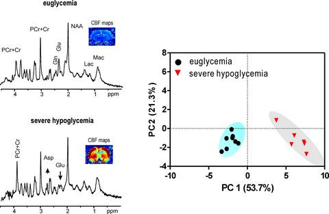
Hypoglycemia is critical condition during diabetic treatment that involves intensive insulin therapy, and it may impair brain function. We aimed to compare cortical responses of three hypoglycemic phases and the restoration of glycemia to control levels after a severe episode in rats using non‐invasive perfusion magnetic resonance (MR) imaging and localized 1H MR spectroscopy. Under light α‐chloralose anesthesia, cortical blood flow (cCBF) was 42 ± 3 ml/100 g/min at euglycemia (~ 5 mM plasma glucose), was not altered at mild hypoglycemia I (42 ± 4 ml/100 g/min, 2–3.5 mM), increased to 60 ± 8 ml/100 g/min under moderate hypoglycemia II (1–2 mM) and amplified to 190 ± 35 ml/100 g/min at severe hypoglycemia III (< 1 mM). 1H MRS revealed metabolic changes at hypoglycemia I without any perfusion alteration. At hypoglycemia III, glutamine and glutamate decreased, whereas aspartate increased. When animals subsequently regained glycemic control, not all metabolites returned to their control levels, for example, glutamine. Meanwhile, ascorbate was increased with amplified hypoglycemic severity, whereas glutathione was reduced; these compounds did not return to normal levels upon the restoration of glycemia. Our study is the first to report cCBF and neurochemical changes in cortex upon five glycemic stages. The cortical responses of different hypoglycemic phases would explain variable neuronal damages after hypoglycemia and might help identify the degrees of hypoglycemic insults and further improve alternative therapies.
中文翻译:

大鼠皮层对急性低血糖的代谢和灌注反应:一种非侵入性磁共振方法。
低血糖症是涉及强化胰岛素治疗的糖尿病治疗中的关键疾病,可能会损害脑功能。我们旨在使用无创灌注磁共振(MR)成像和局部1 H MR谱图比较大鼠严重发作后三个低血糖期的皮质反应和血糖恢复至控制水平。在轻度α-氯海藻糖麻醉下,正常血糖(〜5 mM血浆葡萄糖)时的皮质血流(cCBF)为42±3 ml / 100 g / min,在轻度低血糖I(42±4 ml / 100 g / min)不变(2-3.5 mM),在中度低血糖II(1-2 mM)下增加到60±8 ml / 100 g / min,在严重低血糖III(<1 mM)下放大到190±35 ml / 100 g / min。1个H MRS揭示了低血糖I时的代谢变化,而没有任何灌注改变。在低血糖III期,谷氨酰胺和谷氨酸盐减少,而天冬氨酸增加。当动物随后恢复血糖控制时,并非所有代谢物都恢复到其控制水平,例如谷氨酰胺。同时,随着低血糖严重程度的增加,抗坏血酸增加,而谷胱甘肽减少。这些化合物在恢复血糖后未恢复正常水平。我们的研究是第一个报告在五个血糖阶段皮质中cCBF和神经化学变化的研究。不同的低血糖阶段的皮层反应可以解释低血糖后神经元的可变损伤,并可能有助于确定低血糖损害的程度并进一步改善替代疗法。
更新日期:2020-06-25
中文翻译:
大鼠皮层对急性低血糖的代谢和灌注反应:一种非侵入性磁共振方法。
低血糖症是涉及强化胰岛素治疗的糖尿病治疗中的关键疾病,可能会损害脑功能。我们旨在使用无创灌注磁共振(MR)成像和局部1 H MR谱图比较大鼠严重发作后三个低血糖期的皮质反应和血糖恢复至控制水平。在轻度α-氯海藻糖麻醉下,正常血糖(〜5 mM血浆葡萄糖)时的皮质血流(cCBF)为42±3 ml / 100 g / min,在轻度低血糖I(42±4 ml / 100 g / min)不变(2-3.5 mM),在中度低血糖II(1-2 mM)下增加到60±8 ml / 100 g / min,在严重低血糖III(<1 mM)下放大到190±35 ml / 100 g / min。1个H MRS揭示了低血糖I时的代谢变化,而没有任何灌注改变。在低血糖III期,谷氨酰胺和谷氨酸盐减少,而天冬氨酸增加。当动物随后恢复血糖控制时,并非所有代谢物都恢复到其控制水平,例如谷氨酰胺。同时,随着低血糖严重程度的增加,抗坏血酸增加,而谷胱甘肽减少。这些化合物在恢复血糖后未恢复正常水平。我们的研究是第一个报告在五个血糖阶段皮质中cCBF和神经化学变化的研究。不同的低血糖阶段的皮层反应可以解释低血糖后神经元的可变损伤,并可能有助于确定低血糖损害的程度并进一步改善替代疗法。


 京公网安备 11010802027423号