当前位置:
X-MOL 学术
›
NMR Biomed.
›
论文详情
Our official English website, www.x-mol.net, welcomes your feedback! (Note: you will need to create a separate account there.)
Diffusion tensor magnetic resonance imaging of the postoperative spine with metallic implants.
NMR in Biomedicine ( IF 2.9 ) Pub Date : 2020-04-29 , DOI: 10.1002/nbm.4321 Lian Yang 1 , Yuan Liu 1 , Xiangchuang Kong 1 , Xiaodong Guo 2 , Xiaoming Liu 1 , Qian Qi 3 , Jiazheng Wang 3
NMR in Biomedicine ( IF 2.9 ) Pub Date : 2020-04-29 , DOI: 10.1002/nbm.4321 Lian Yang 1 , Yuan Liu 1 , Xiangchuang Kong 1 , Xiaodong Guo 2 , Xiaoming Liu 1 , Qian Qi 3 , Jiazheng Wang 3
Affiliation
|
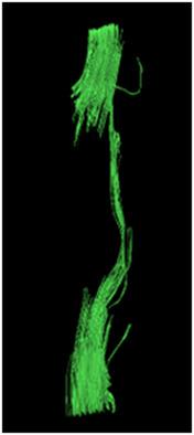
There has been a growing need to understand the mechanism of development of acute spinal cord injury (SCI) and to optimize treatment. The paramagnetic nature of metallic implants has hampered the application of diffusion tensor imaging (DTI) in postsurgical SCI monitoring. We describe here a successful implementation of spinal DTI in postsurgical SCI patients. Data were acquired using a single‐shot turbo‐spin‐echo sequence, where an extra gradient is applied before the refocusing pulse train to eliminate contributions from the non‐Carr‐Purcell‐Meiboom‐Gill components following a diffusion preparation block where a single‐spin echo scheme is deployed. The DTI images were acquired in axial orientation with a 2 x 2 x 4 mm3 resolution and a total of 18 slices. Diffusion gradients were applied in six directions with b values of 0 and 600 seconds/mm2. The whole scan took ~10 minutes. The sequence was compared with SENSE‐DW‐EPI and ZOOM‐DW‐EPI on a phantom, eight patients with either anterior or posterior titanium alloy implants, and a pork loin with a similar implant. The protocol resulted in dramatically reduced geometric distortions compared with routine imaging sequences, however, the SNR efficiency was compromised. The spinal cord signal displacement was 0.68±1.00 mm (mean±SD, n = 8) for the proposed protocol, and 5.14±3.07 and 2.82±1.60 mm for the SENSE‐DW‐EPI and ZOOM‐DW‐EPI sequences, respectively. Fiber tracking was achieved in the presence of implants, which in one case was accompanied by central spinal cord caviation. Mathematical analysis concluded that the proposed protocol would be generally applicable in the spinal cord when the titanium alloy implant is ~15 mm away (<0.5 kHz B0 field drift). The protocol described is capable of DTI in postsurgery SCI patients with metallic implants at sufficient resolution and SNR.
中文翻译:

术后脊柱金属植入物的弥散张量磁共振成像。
人们越来越需要了解急性脊髓损伤 (SCI) 的发展机制并优化治疗。金属植入物的顺磁性阻碍了扩散张量成像 (DTI) 在术后 SCI 监测中的应用。我们在这里描述了脊柱 DTI 在术后 SCI 患者中的成功实施。使用单次涡轮自旋回波序列获取数据,在重聚焦脉冲序列之前应用额外的梯度,以消除来自非 Carr-Purcell-Meiboom-Gill 组件的贡献,在扩散准备块之后,单次部署了自旋回波方案。DTI 图像是在轴向方向上用 2 x 2 x 4 mm 3分辨率和总共 18 个切片。在六个方向上应用扩散梯度,b 值为 0 和 600 秒/mm 2。整个扫描大约需要 10 分钟。该序列与 SENSE-DW-EPI 和 ZOOM-DW-EPI 在一个体模、8 名具有前部或后部钛合金植入物的患者以及具有类似植入物的猪里脊肉上进行比较。与常规成像序列相比,该协议显着减少了几何失真,但是,SNR 效率受到了影响。拟议协议的脊髓信号位移为 0.68 ± 1.00 毫米(平均值± SD,n = 8),5.14 ± 3.07 和 2.82 ±SENSE-DW-EPI 和 ZOOM-DW-EPI 序列分别为 1.60 mm。在存在植入物的情况下实现了纤维追踪,其中一种情况是伴有中央脊髓空洞。数学分析得出结论,当钛合金植入物距离约 15 毫米(<0.5 kHz B 0场漂移)时,所提出的协议将普遍适用于脊髓。所描述的协议能够在具有足够分辨率和 SNR 的金属植入物的术后 SCI 患者中进行 DTI。
更新日期:2020-07-08
中文翻译:
术后脊柱金属植入物的弥散张量磁共振成像。
人们越来越需要了解急性脊髓损伤 (SCI) 的发展机制并优化治疗。金属植入物的顺磁性阻碍了扩散张量成像 (DTI) 在术后 SCI 监测中的应用。我们在这里描述了脊柱 DTI 在术后 SCI 患者中的成功实施。使用单次涡轮自旋回波序列获取数据,在重聚焦脉冲序列之前应用额外的梯度,以消除来自非 Carr-Purcell-Meiboom-Gill 组件的贡献,在扩散准备块之后,单次部署了自旋回波方案。DTI 图像是在轴向方向上用 2 x 2 x 4 mm 3分辨率和总共 18 个切片。在六个方向上应用扩散梯度,b 值为 0 和 600 秒/mm 2。整个扫描大约需要 10 分钟。该序列与 SENSE-DW-EPI 和 ZOOM-DW-EPI 在一个体模、8 名具有前部或后部钛合金植入物的患者以及具有类似植入物的猪里脊肉上进行比较。与常规成像序列相比,该协议显着减少了几何失真,但是,SNR 效率受到了影响。拟议协议的脊髓信号位移为 0.68 ± 1.00 毫米(平均值± SD,n = 8),5.14 ± 3.07 和 2.82 ±SENSE-DW-EPI 和 ZOOM-DW-EPI 序列分别为 1.60 mm。在存在植入物的情况下实现了纤维追踪,其中一种情况是伴有中央脊髓空洞。数学分析得出结论,当钛合金植入物距离约 15 毫米(<0.5 kHz B 0场漂移)时,所提出的协议将普遍适用于脊髓。所描述的协议能够在具有足够分辨率和 SNR 的金属植入物的术后 SCI 患者中进行 DTI。


 京公网安备 11010802027423号