当前位置:
X-MOL 学术
›
Hum. Brain Mapp.
›
论文详情
Our official English website, www.x-mol.net, welcomes your feedback! (Note: you will need to create a separate account there.)
From a deep learning model back to the brain-Identifying regional predictors and their relation to aging.
Human Brain Mapping ( IF 4.8 ) Pub Date : 2020-04-22 , DOI: 10.1002/hbm.25011 Gidon Levakov 1, 2 , Gideon Rosenthal 1, 2 , Ilan Shelef 2, 3 , Tammy Riklin Raviv 2, 4 , Galia Avidan 1, 2, 5
Human Brain Mapping ( IF 4.8 ) Pub Date : 2020-04-22 , DOI: 10.1002/hbm.25011 Gidon Levakov 1, 2 , Gideon Rosenthal 1, 2 , Ilan Shelef 2, 3 , Tammy Riklin Raviv 2, 4 , Galia Avidan 1, 2, 5
Affiliation
|
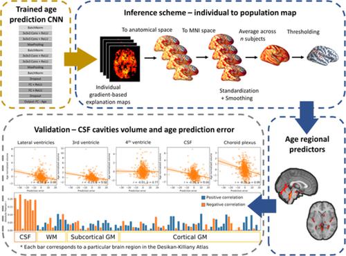
We present a Deep Learning framework for the prediction of chronological age from structural magnetic resonance imaging scans. Previous findings associate increased brain age with neurodegenerative diseases and higher mortality rates. However, the importance of brain age prediction goes beyond serving as biomarkers for neurological disorders. Specifically, utilizing convolutional neural network (CNN) analysis to identify brain regions contributing to the prediction can shed light on the complex multivariate process of brain aging. Previous work examined methods to attribute pixel/voxel‐wise contributions to the prediction in a single image, resulting in “explanation maps” that were found noisy and unreliable. To address this problem, we developed an inference scheme for combining these maps across subjects, thus creating a population‐based, rather than a subject‐specific map. We applied this method to a CNN ensemble trained on predicting subjects' age from raw T1 brain images in a lifespan sample of 10,176 subjects. Evaluating the model on an untouched test set resulted in mean absolute error of 3.07 years and a correlation between chronological and predicted age of r = 0.98. Using the inference method, we revealed that cavities containing cerebrospinal fluid, previously found as general atrophy markers, had the highest contribution for age prediction. Comparing maps derived from different models within the ensemble allowed to assess differences and similarities in brain regions utilized by the model. We showed that this method substantially increased the replicability of explanation maps, converged with results from voxel‐based morphometry age studies and highlighted brain regions whose volumetric variability correlated the most with the prediction error.
中文翻译:

从深度学习模型回到大脑——识别区域预测因子及其与衰老的关系。
我们提出了一个深度学习框架,用于根据结构磁共振成像扫描预测实际年龄。先前的研究结果将大脑年龄的增加与神经退行性疾病和更高的死亡率联系起来。然而,大脑年龄预测的重要性不仅仅是作为神经系统疾病的生物标志物。具体来说,利用卷积神经网络(CNN)分析来识别有助于预测的大脑区域可以揭示大脑衰老的复杂多变量过程。之前的工作研究了将像素/体素方面的贡献归因于单个图像中的预测的方法,导致发现噪声和不可靠的“解释图”。为了解决这个问题,我们开发了一种推理方案,用于将这些跨主题的地图组合起来,从而创建基于人群的地图,而不是特定于主题的地图。我们将此方法应用于 CNN 集合,该集合经过训练,可以根据 10,176 名受试者的生命周期样本中的原始 T1 大脑图像来预测受试者的年龄。在未受影响的测试集上评估模型的结果是平均绝对误差为 3.07 年,实际年龄和预测年龄之间的相关性为r = 0.98。使用推理方法,我们发现含有脑脊液的空洞(以前被发现作为一般萎缩标志物)对年龄预测的贡献最大。比较从整体中不同模型得出的地图可以评估模型所使用的大脑区域的差异和相似性。我们表明,这种方法大大提高了解释图的可重复性,与基于体素的形态测量年龄研究的结果相一致,并突出显示了体积变异性与预测误差最相关的大脑区域。
更新日期:2020-04-22
中文翻译:
从深度学习模型回到大脑——识别区域预测因子及其与衰老的关系。
我们提出了一个深度学习框架,用于根据结构磁共振成像扫描预测实际年龄。先前的研究结果将大脑年龄的增加与神经退行性疾病和更高的死亡率联系起来。然而,大脑年龄预测的重要性不仅仅是作为神经系统疾病的生物标志物。具体来说,利用卷积神经网络(CNN)分析来识别有助于预测的大脑区域可以揭示大脑衰老的复杂多变量过程。之前的工作研究了将像素/体素方面的贡献归因于单个图像中的预测的方法,导致发现噪声和不可靠的“解释图”。为了解决这个问题,我们开发了一种推理方案,用于将这些跨主题的地图组合起来,从而创建基于人群的地图,而不是特定于主题的地图。我们将此方法应用于 CNN 集合,该集合经过训练,可以根据 10,176 名受试者的生命周期样本中的原始 T1 大脑图像来预测受试者的年龄。在未受影响的测试集上评估模型的结果是平均绝对误差为 3.07 年,实际年龄和预测年龄之间的相关性为r = 0.98。使用推理方法,我们发现含有脑脊液的空洞(以前被发现作为一般萎缩标志物)对年龄预测的贡献最大。比较从整体中不同模型得出的地图可以评估模型所使用的大脑区域的差异和相似性。我们表明,这种方法大大提高了解释图的可重复性,与基于体素的形态测量年龄研究的结果相一致,并突出显示了体积变异性与预测误差最相关的大脑区域。


 京公网安备 11010802027423号