当前位置:
X-MOL 学术
›
Eur. J. Nerosci.
›
论文详情
Our official English website, www.x-mol.net, welcomes your feedback! (Note: you will need to create a separate account there.)
Midbrain control of breathing and blood pressure: The role of periaqueductal gray matter and mesencephalic collicular neuronal microcircuit oscillators.
European Journal of Neroscience ( IF 3.4 ) Pub Date : 2020-03-29 , DOI: 10.1111/ejn.14727 Michael George Zaki Ghali 1, 2, 3
European Journal of Neroscience ( IF 3.4 ) Pub Date : 2020-03-29 , DOI: 10.1111/ejn.14727 Michael George Zaki Ghali 1, 2, 3
Affiliation

|
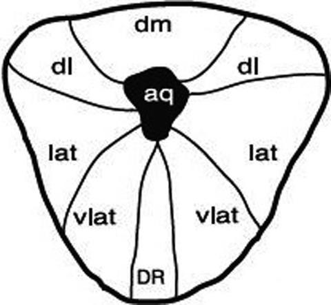
Neural circuitry residing within the medullary ventral respiratory column nuclei and dorsal respiratory group interact with the Kölliker‐Fuse and medial parabrachial nuclei to generate the core breathing rhythm and pattern during resting conditions. Triphasic eupnea consists of inspiratory [I], post‐inspiratory [post‐I], and late‐expiratory [E2] phases. Mesencephalic zones exert modulatory influences upon respiratory rhythm‐generating circuitry, sympathetic oscillators, and parasympathetic nuclei. The earliest evidence supporting the existence of midbrain control of breathing derives from studies conducted by Martin and Booker in 1878. These authors demonstrated electrical stimulation of the deep layers of the mesencephalic colliculi in the rabbit augmented ventilation and sequentially elicited chest wall tremors and tetany. Investigations performed during the past several decades would demonstrate stimlation of distributed zones within the midbrain reticular formation elicits starkly disparate effects upon respiratory phase switching. Schmid, Böhmer, and Fallert demonstrated electrical stimulation of the nucleus rubre and emanating axon bundles alternately elicits or inhibits the activity of medullary expiratory‐ or inspiratory‐related units and phrenic nerve discharge with differential latency. A series of studies would later indicate the red nucleus mediates hypoxic ventilatory depression. Periaqueductal gray matter neurons exhibit extensive afferent and efferent interconnectivity with suprabulbar, brainstem, and spinal cord zones aptly positioning these units to modulate breathing, autonomic outflow, nociception locomotion, micturtion, and sexual behavior. Experimental stimulatory activation of the tectal colliculi and periaqueductal gray matter via electrical current or glutamate, D,L‐homocysteinic acid, or bicuculline microinjections coordinately modulates neuromotor inspiratory bursting frequency and amplitude and discharge of pre‐Bötzinger complex, ventrolateral medullary late‐I and post‐I, and ventrolateral nucleus tractus solitarius decrementing early‐I and augmenting and decrementing late‐I neurons, elicits expiratory outflow and vocalization, and blunt the Hering‐Breuer reflex in unanesthetzed decerebrate and anesthetized preprations of the cat and rat. Stimulation of the mesencephalic colliuli or dorsal divisions of the PAG potently amplifes renal sympathetic neural efferent activity, dynamic arterial pressure magnitude, and myocardial contraction frequency and elicits various behavioral defense responses. Elicited physiological effects exhibit extensive locoregional heterogeneity and variably enlist requisite contributions from the dorsomedial hypothalamus and/or lateral parabrachial nuclei. Stimulation of the dorsal mesencephalon occasionally elicits dynamic increases of arterial pressure magnitude exhibiting prominent oscillatory variability coherent with phrenic nerve discharge, perhaps by generating intra‐neuraxial hysteresis, serving to intermittently deliver blood to organ vascular beds under high pressure in order to prevent organ edema, microcirculatory dysfunction, and downregulation of vascular smooth muscle alpha adrenergic receptors. Chemosensitive mesencephalic caudal raphé units and projections of hypoxia‐sensitive units in the caudal hypothalamus to the periaqueductal gray matter may imply the existence of a diencephalo‐smesencephalic chemosensitive network modulating breathing and sympathetic discharge.
中文翻译:

呼吸和血压的中脑控制:导水管周围灰质和中脑颈神经元微电路振荡器的作用。
位于延髓腹侧呼吸柱核和背侧呼吸群内的神经回路与 Kölliker-Fuse 和内侧臂旁核相互作用,在休息条件下产生核心呼吸节律和模式。三相呼吸暂停由吸气 [I]、吸气后 [post-I] 和呼气后期 [E2] 阶段组成。中脑区对呼吸节律产生回路、交感神经振荡器和副交感神经核产生调节影响。支持中脑呼吸控制存在的最早证据来自 Martin 和 Booker 于 1878 年进行的研究。这些作者证明了在兔子增强通气中对中脑丘深层进行电刺激并依次引起胸壁震颤和手足抽搐。在过去几十年中进行的调查表明,对中脑网状结构内分布区域的刺激对呼吸相位转换产生了截然不同的影响。Schmid、Böhmer 和 Fallert 证明了电刺激红核和发出的轴突束交替引发或抑制延髓呼气或吸气相关单位和膈神经放电的活动,具有不同的潜伏期。后来的一系列研究表明,红核介导了缺氧通气抑制。导水管周围灰质神经元与球上、脑干和脊髓区域表现出广泛的传入和传出互连,这些区域恰当地定位这些单元以调节呼吸、自主神经流出、伤害感受运动、排尿和性行为。通过电流或谷氨酸盐、D、L-高半胱氨酸或荷包牡丹碱显微注射对顶盖丘和导水管周围灰质的实验刺激激活协调调节神经运动吸气爆发频率和振幅以及前 Bötzinger 复合体、腹外侧延髓 I 和后期的放电‐I 和腹外侧孤束核减少早期 I 和增加和减少晚期 I 神经元,引起呼气流出和发声,并使猫和大鼠的未麻醉去脑和麻醉制剂中的 Hering-Breuer 反射减弱。刺激 PAG 的中脑颈或背侧分裂有效地增强肾交感神经传出活动、动态动脉压大小、和心肌收缩频率并引发各种行为防御反应。引发的生理效应表现出广泛的局部异质性,并可变地从背内侧下丘脑和/或外侧臂旁核中获得必要的贡献。中脑背侧的刺激偶尔会引起动脉压的动态增加,表现出与膈神经放电相关的显着振荡变异性,可能是通过产生神经轴内滞后,在高压下间歇性地将血液输送到器官血管床以防止器官水肿,微循环功能障碍和血管平滑肌α肾上腺素能受体的下调。
更新日期:2020-03-29
中文翻译:

呼吸和血压的中脑控制:导水管周围灰质和中脑颈神经元微电路振荡器的作用。
位于延髓腹侧呼吸柱核和背侧呼吸群内的神经回路与 Kölliker-Fuse 和内侧臂旁核相互作用,在休息条件下产生核心呼吸节律和模式。三相呼吸暂停由吸气 [I]、吸气后 [post-I] 和呼气后期 [E2] 阶段组成。中脑区对呼吸节律产生回路、交感神经振荡器和副交感神经核产生调节影响。支持中脑呼吸控制存在的最早证据来自 Martin 和 Booker 于 1878 年进行的研究。这些作者证明了在兔子增强通气中对中脑丘深层进行电刺激并依次引起胸壁震颤和手足抽搐。在过去几十年中进行的调查表明,对中脑网状结构内分布区域的刺激对呼吸相位转换产生了截然不同的影响。Schmid、Böhmer 和 Fallert 证明了电刺激红核和发出的轴突束交替引发或抑制延髓呼气或吸气相关单位和膈神经放电的活动,具有不同的潜伏期。后来的一系列研究表明,红核介导了缺氧通气抑制。导水管周围灰质神经元与球上、脑干和脊髓区域表现出广泛的传入和传出互连,这些区域恰当地定位这些单元以调节呼吸、自主神经流出、伤害感受运动、排尿和性行为。通过电流或谷氨酸盐、D、L-高半胱氨酸或荷包牡丹碱显微注射对顶盖丘和导水管周围灰质的实验刺激激活协调调节神经运动吸气爆发频率和振幅以及前 Bötzinger 复合体、腹外侧延髓 I 和后期的放电‐I 和腹外侧孤束核减少早期 I 和增加和减少晚期 I 神经元,引起呼气流出和发声,并使猫和大鼠的未麻醉去脑和麻醉制剂中的 Hering-Breuer 反射减弱。刺激 PAG 的中脑颈或背侧分裂有效地增强肾交感神经传出活动、动态动脉压大小、和心肌收缩频率并引发各种行为防御反应。引发的生理效应表现出广泛的局部异质性,并可变地从背内侧下丘脑和/或外侧臂旁核中获得必要的贡献。中脑背侧的刺激偶尔会引起动脉压的动态增加,表现出与膈神经放电相关的显着振荡变异性,可能是通过产生神经轴内滞后,在高压下间歇性地将血液输送到器官血管床以防止器官水肿,微循环功能障碍和血管平滑肌α肾上腺素能受体的下调。


 京公网安备 11010802027423号