当前位置:
X-MOL 学术
›
J. Neurosci. Res.
›
论文详情
Our official English website, www.x-mol.net, welcomes your feedback! (Note: you will need to create a separate account there.)
Advancing zebrafish as a model for studying developmental neurotoxicology.
Journal of Neuroscience Research ( IF 4.2 ) Pub Date : 2020-03-30 , DOI: 10.1002/jnr.24621 Nathan R Martin 1, 2, 3 , Jessica S Plavicki 1, 2, 3
Journal of Neuroscience Research ( IF 4.2 ) Pub Date : 2020-03-30 , DOI: 10.1002/jnr.24621 Nathan R Martin 1, 2, 3 , Jessica S Plavicki 1, 2, 3
Affiliation
|
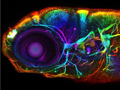
The cover photo shows the developing zebrafish nervous system at 5 days post-fertilization. Axon tracts are labeled with an anti-acetylated alpha tubulin antibody. The image, which was acquired on a Zeiss LSM 880 confocal microscope, is a maximum intensity projection of a z-stack that has been color-coded for depth. Major brain regions such as the olfactory bulb, forebrain, habenula, optic tectum, cerebellum, hindbrain, and eye are identifiable. This image is part of a study (Plavicki Lab, Brown University) focused on understanding the impact of toxicant exposures on brain development and activity with the goal of identifyingenvironmental factors that contribute to the etiology of neurodevelopmental disorders.
中文翻译:

推进斑马鱼作为研究发育神经毒理学的模型。
封面照片显示了受精后 5 天发育中的斑马鱼神经系统。轴突束用抗乙酰化α微管蛋白抗体标记。该图像是在 Zeiss LSM 880 共聚焦显微镜上采集的,是 z 堆栈的最大强度投影,已对深度进行颜色编码。主要的大脑区域,如嗅球、前脑、缰核、视顶盖、小脑、后脑和眼睛是可识别的。该图像是一项研究(布朗大学普拉维奇实验室)的一部分,该研究专注于了解毒物暴露对大脑发育和活动的影响,目的是确定导致神经发育障碍病因的环境因素。
更新日期:2020-03-30
中文翻译:
推进斑马鱼作为研究发育神经毒理学的模型。
封面照片显示了受精后 5 天发育中的斑马鱼神经系统。轴突束用抗乙酰化α微管蛋白抗体标记。该图像是在 Zeiss LSM 880 共聚焦显微镜上采集的,是 z 堆栈的最大强度投影,已对深度进行颜色编码。主要的大脑区域,如嗅球、前脑、缰核、视顶盖、小脑、后脑和眼睛是可识别的。该图像是一项研究(布朗大学普拉维奇实验室)的一部分,该研究专注于了解毒物暴露对大脑发育和活动的影响,目的是确定导致神经发育障碍病因的环境因素。


 京公网安备 11010802027423号